Application
IP analysis was performed using 500 ug of HeLa lysate and 3 ug of PAX7 antibody. The precipitate was detected by the same antibody with EasyBlot as the secondary antibody.Application
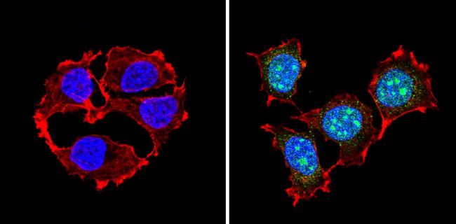
ICC/IF analysis of MCF7 cells with (right) or without (left) PAX7 antibody at a dilution of 1:100 (green). F-actin (red) was stained with a flourescent red phalloidin and nuclei (blue) were stained with Hoechst or DAPI. Images were taken at a magnification of 60x.Application
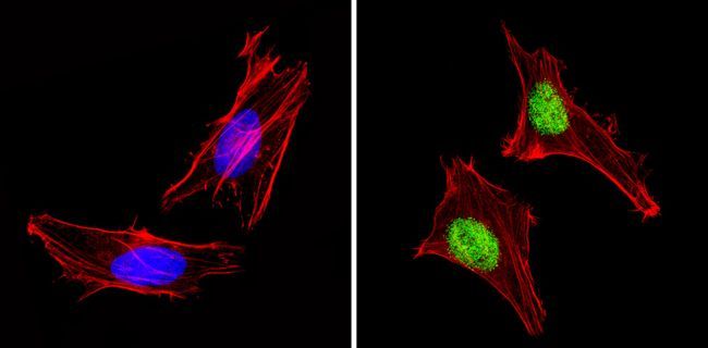
ICC/IF analysis of HeLa cells with (right) or without (left) PAX7 antibody at a dilution of 1:100 (green). F-actin (red) was stained with a flourescent red phalloidin and nuclei (blue) were stained with Hoechst or DAPI. Images were taken at a magnification of 60x.Application
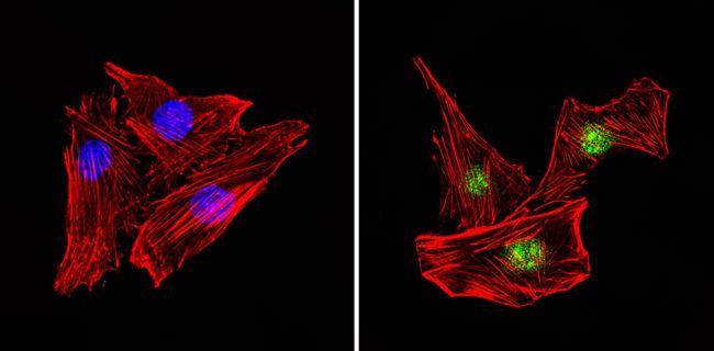
ICC/IF analysis of C2C12 cells with (right) or without (left) PAX7 antibody at a dilution of 1:100 (green). F-actin (red) was stained with a flourescent red phalloidin and nuclei (blue) were stained with Hoechst or DAPI. Images were taken at a magnification of 60x.Application
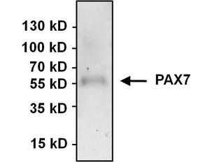
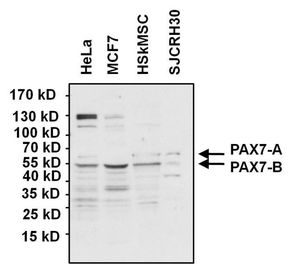
WB analysis of various cell lysates (50 ug per lane) using PAX7 antibody at a dilution of 1:1000.Application
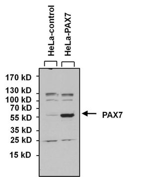
WB analysis of lysates from HeLa cells overexpressing PAX7 (right) or empty vector control (left) using PAX7 antibody at a dilution of 1:1000.| Product Name | PAX7 antibody |
|---|---|
| Antibody Type | Primary Antibodies |
| Product description | Rabbit polyclonal antibody to PAX7This gene is a member of the paired box (PAX) family of transcription factors. Members of this gene family typically contain a paired box domain, an octapeptide, and a paired-type homeodomain. These genes play critical roles during fetal development and cancer growth. The specific function of the paired box 7 gene is unknown but speculated to involve tumor suppression since fusion of this gene with a forkhead domain family member has been associated with alveolar rhabdomyosarcoma. Three transcript variants encoding different isoforms have been found for this gene. [provided by RefSeq, Sep 2008] |
| Immunogen | Purified internal fragment of human recombinant PAX7 expressed in E. coli. |
| Clonality | Polyclonal |
|---|---|
| Isotype | IgG |
| Host Species | Rabbit |
| Tested Applications | ICC/IFIPWB |
| ICC/IF:1:200~1:500 IP:1:100~1:500 WB:1:500~1:2000 Notes:Optimal dilutions/concentrations should be determined by the researcher. |
|
| Species Reactivity | HumanMouse |
| Concentration | 1mg/ml |
| Purification | Protein G |
| Gene Synonyms | paired box 7 PAX7B RMS2 HUP1 |
|---|---|
| Target | PAX7 |
Application
IP analysis was performed using 500 ug of HeLa lysate and 3 ug of PAX7 antibody. The precipitate was detected by the same antibody with EasyBlot as the secondary antibody.
Application
ICC/IF analysis of MCF7 cells with (right) or without (left) PAX7 antibody at a dilution of 1:100 (green). F-actin (red) was stained with a flourescent red phalloidin and nuclei (blue) were stained with Hoechst or DAPI. Images were taken at a magnification of 60x.
Application
ICC/IF analysis of HeLa cells with (right) or without (left) PAX7 antibody at a dilution of 1:100 (green). F-actin (red) was stained with a flourescent red phalloidin and nuclei (blue) were stained with Hoechst or DAPI. Images were taken at a magnification of 60x.
Application
ICC/IF analysis of C2C12 cells with (right) or without (left) PAX7 antibody at a dilution of 1:100 (green). F-actin (red) was stained with a flourescent red phalloidin and nuclei (blue) were stained with Hoechst or DAPI. Images were taken at a magnification of 60x.
Application
WB analysis of various cell lysates (50 ug per lane) using PAX7 antibody at a dilution of 1:1000.
Application
WB analysis of lysates from HeLa cells overexpressing PAX7 (right) or empty vector control (left) using PAX7 antibody at a dilution of 1:1000.| Application Notes | ICC/IF:1:200~1:500 IP:1:100~1:500 WB:1:500~1:2000 Notes:Optimal dilutions/concentrations should be determined by the researcher. |
|---|
| Form | Liquid |
|---|---|
| Storage Instructions | -20° C |
| Storage Buffer | PBS with 1mg/ml BSA, 30% glycerol |


