Application
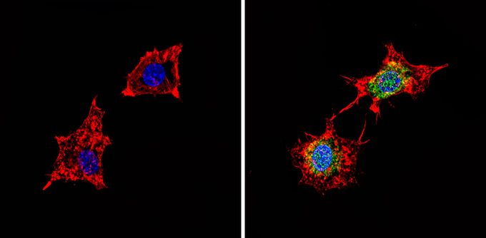
ICC/IF analysis of BAEC cells with (right) or without (left) using PDE6 delta antibody at a dilution of 1:100 (green). F-actin (red) was stained with a flourescent red phalloidin and nuclei (blue) were stained with DAPI. Images were taken at a magnification of 60x.Application
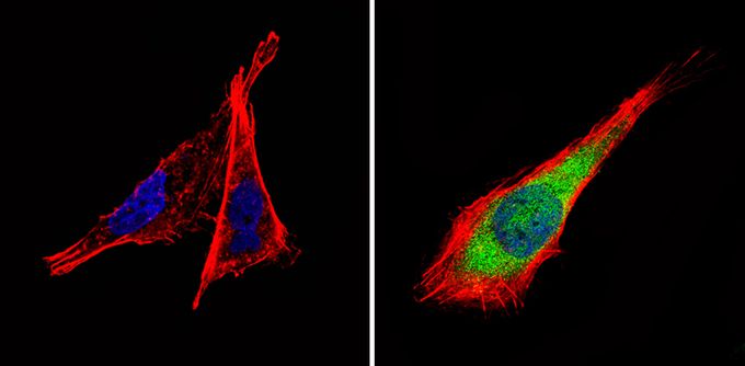
ICC/IF analysis of B-3 cells with (right) or without (left) PDE6 delta antibody at a dilution of 1:100 (green). F-actin (red) was stained with a flourescent red phalloidin and nuclei (blue) were stained with DAPI. Images were taken at a magnification of 60x.| Product Name | PDE6D antibody |
|---|---|
| Antibody Type | Primary Antibodies |
| Product description | Rabbit Polyclonal antibody to PDE6DPrBP/delta, previously known as PDE6/delta, is the effector enzyme in the G protein-mediated signal transduction cascade in the visual system. There are five different subunits consisting of rod and cone specific catalytic subunits: alpha (Cone), alpha (Rod), and beta (Rod), the inhibitory subunit gamma, and subunit delta of unknown function (which likely interacts with many other proteins besides the PDE6 family). PRBP/delta has been implicated in the transport and membrane targeting of prenylated proteins (including PDE6) from their site of synthesis in the inner segment to their final destination in the outer segment of rods and cones (Norton et. al., 2005). |
| Immunogen | Synthetic Peptides: M(1) S A K D E R A R (D/E) I L R G F K(16) C |
| Clonality | Polyclonal |
|---|---|
| Host Species | Rabbit |
| Tested Applications | ICC/IFIHCIHC-FWB |
| ICC/IF: 1:10 - 1:200: |
|
| Species Reactivity | HumanMousePigSheepBovine |
| Concentration | 1mg/ml (Please refer to the vial label for the specific concentration) |
| Purification | Affinity purified |
| Gene Synonyms | Pde6d AI841218 PrBP/delta phosphodiesterase 6D cGMP-specific rod delta |
|---|---|
| Target | PDE6 delta |
Application
ICC/IF analysis of BAEC cells with (right) or without (left) using PDE6 delta antibody at a dilution of 1:100 (green). F-actin (red) was stained with a flourescent red phalloidin and nuclei (blue) were stained with DAPI. Images were taken at a magnification of 60x.
Application
ICC/IF analysis of B-3 cells with (right) or without (left) PDE6 delta antibody at a dilution of 1:100 (green). F-actin (red) was stained with a flourescent red phalloidin and nuclei (blue) were stained with DAPI. Images were taken at a magnification of 60x.| Application Notes | ICC/IF: 1:10 - 1:200: |
|---|
| Form | Liquid |
|---|---|
| Storage Instructions | Keep as concentrated solution. Aliquot and store at -20ºC or below. Avoid multiple freeze-thaw cycles. |
| Storage Buffer | PBS containing 1 mg/ml BSA and 0.05% sodium azide. |


