Application
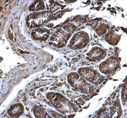
PDI antibody [N1N3] detects PDI protein at cytosol and membrane on mouse colon by immunohistochemical analysis. Sample: Paraffin-embedded mouse colon. PDI antibody [N1N3] dilution: 1:500.Application
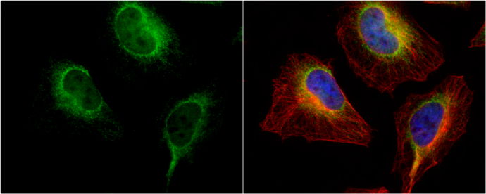
PDI antibody [N1N3] detects PDI protein at endoplasmic reticulum by immunofluorescent analysis. Sample: HeLa cells were fixed in 4% paraformaldehyde at RT for 15 min. Green: PDI protein stained by PDI antibody [N1N3] diluted at 1:100. Red: phalloidin, a cytoskeleton marker, stained by phalloidin (invitrogen, A12380) diluted at 1:200. Blue: Hoechst 33342 staining.Application
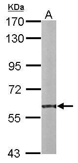
Sample (20 ug of whole cell lysate) A: mouse liver 7.5% SDS PAGE diluted at 1:10000Application
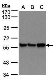
Sample(30 μg of whole cell lysate) A:293T B:A431 C:H1299 7.5% SDS PAGE diluted at 1:500Application
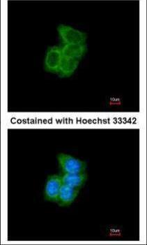
Immunofluorescence analysis of methanol-fixed A431, using PDI antibody at 1:200 dilution.Application
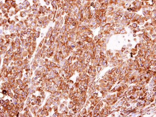
Immunohistochemical analysis of paraffin-embedded H520 xenograft, using PDI antibody at 1:100 dilution.| Product Name | PDI antibody |
|---|---|
| Antibody Type | Primary Antibodies |
| Product description | Rabbit Polyclonal antibody to PDI (prolyl 4-hydroxylase, beta polypeptide)This gene encodes the beta subunit of prolyl 4-hydroxylase, a highly abundant multifunctional enzyme that belongs to the protein disulfide isomerase family. When present as a tetramer consisting of two alpha and two beta subunits, this enzyme is involved in hydroxylation of prolyl residues in preprocollagen. This enzyme is also a disulfide isomerase containing two thioredoxin domains that catalyze the formation, breakage and rearrangement of disulfide bonds. Other known functions include its ability to act as a chaperone that inhibits aggregation of misfolded proteins in a concentration-dependent manner, its ability to bind thyroid hormone, its role in both the influx and efflux of S-nitrosothiol-bound nitric oxide, and its function as a subunit of the microsomal triglyceride transfer protein complex. [provided by RefSeq] |
| Immunogen | Recombinant protein encompassing a sequence within the center region of human PDI. The exact sequence is proprietary. |
| Clonality | Polyclonal |
|---|---|
| Isotype | IgG |
| Host Species | Rabbit |
| Tested Applications | EMICC/IFIHC-PWB |
| *Optimal dilutions/concentrations should be determined by the researcher.
: |
|
| Species Reactivity | HumanMouse |
| Predicted Reactivity | Rat, Rabbit, Chicken, Chimpanzee, BovineRat (92%), Rabbit (91%), Chicken (83%), Chimpanzee (100%), Bovine (93%) |
| Concentration | 1mg/ml (Please refer to the vial label for the specific concentration) |
| Purification | Affinity purified |
| Gene Synonyms | DSI antibody ERBA2L antibody GIT antibody P4Hbeta antibody PDI antibody PDIA1 antibody PHDB antibody PO4DB antibody PO4HB antibody PROHB antibody P4HB antibody cellular thyroid hormone-binding protein antibody protein disulfide-isomerase antibo |
|---|---|
| Molecular Weight(MW) | 57 kDa (note) |
| Cellular Localization | Endoplasmic reticulum lumen , Melanosome , Cell membrane; Peripheral membrane protein (Potential) |
Application
PDI antibody [N1N3] detects PDI protein at cytosol and membrane on mouse colon by immunohistochemical analysis. Sample: Paraffin-embedded mouse colon. PDI antibody [N1N3] dilution: 1:500.
Application
PDI antibody [N1N3] detects PDI protein at endoplasmic reticulum by immunofluorescent analysis. Sample: HeLa cells were fixed in 4% paraformaldehyde at RT for 15 min. Green: PDI protein stained by PDI antibody [N1N3] diluted at 1:100. Red: phalloidin, a cytoskeleton marker, stained by phalloidin (invitrogen, A12380) diluted at 1:200. Blue: Hoechst 33342 staining.
Application
Sample (20 ug of whole cell lysate) A: mouse liver 7.5% SDS PAGE diluted at 1:10000
Application
Sample(30 μg of whole cell lysate) A:293T B:A431 C:H1299 7.5% SDS PAGE diluted at 1:500
Application
Immunofluorescence analysis of methanol-fixed A431, using PDI antibody at 1:200 dilution.
Application
Immunohistochemical analysis of paraffin-embedded H520 xenograft, using PDI antibody at 1:100 dilution.| Positive Control | 293T , A431 , H1299 , HeLaS3 , HepG2 , Molt-4 , Raji , mouse liver |
|---|---|
| Application Notes | *Optimal dilutions/concentrations should be determined by the researcher.
: |
| Form | Liquid |
|---|---|
| Storage Instructions | Keep as concentrated solution. Aliquot and store at -20ºC or below. Avoid multiple freeze-thaw cycles. |
| Storage Buffer | 0.1M Tris, 0.1M Glycine, 10% Glycerol (pH7). 0.01% Thimerosal was added as a preservative. |


