Application
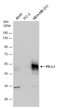
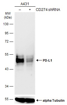
Various whole cell extracts (30 µg) were separated by 10% SDS-PAGE, and the membrane was blotted with PD-L1 antibody diluted at 1:2000.Application
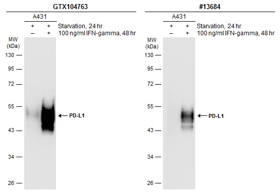
Untreated (–) and treated (+) A431 whole cell extracts (30 µg) were separated by 10% SDS-PAGE, and the membranes were blotted with PD-L1 antibody diluted at 1:6000 and competitor's antibody (CST#13684) diluted at 1:500. The HRP-conjugated anti-rabbit IgG antibody was used to detect the primary antibody.Application
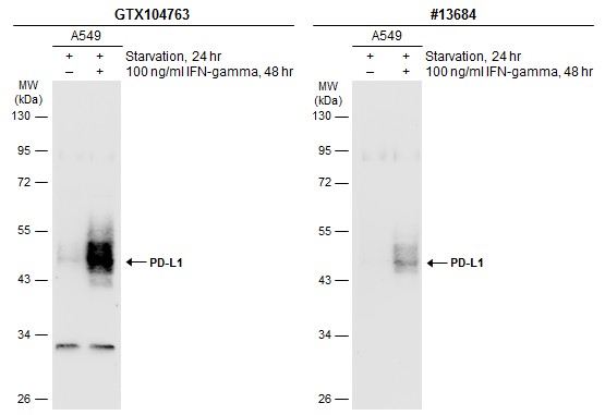
Untreated (–) and treated (+) A549 whole cell extracts (30 µg) were separated by 10% SDS-PAGE, and the membranes were blotted with PD-L1 antibody diluted at 1:3000 and competitor's antibody (CST#13684) diluted at 1:500. The HRP-conjugated anti-rabbit IgG antibody was used to detect the primary antibody.Application
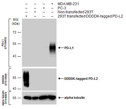
Various whole cell extracts were separated by 10% SDS-PAGE, and the membranes were blotted with PD-L1 antibody diluted at 1:3000 and with DDDDK tag antibody diluted at 1:3000 to detect DDDDK-tagged PD-L2. The HRP-conjugated anti-rabbit IgG antibody was used to detect the primary antibody.Application
Non-transfected (–) and transfected (+) A431 whole cell extracts (30 µg) were separated by 10% SDS-PAGE, and the membrane was blotted with PD-L1 antibody diluted at 1:5000. The HRP-conjugated anti-rabbit IgG antibody was used to detect the primary antibody.Application
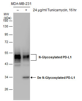
Untreated (–) and treated (+) MDA-MB-231 whole cell extracts (30 µg) were separated by 10% SDS-PAGE, and the membrane was blotted with PD-L1 antibody diluted at 1:5000.Application
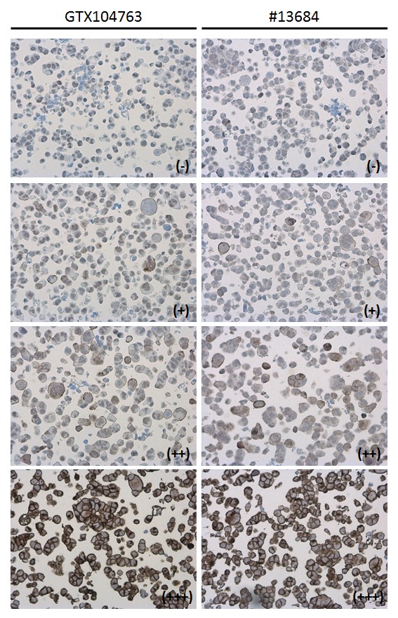
PD-L1 antibody detects PD-L1 protein at cell membrane in PD-L1 protein-expressing cell lines by immunohistochemical analysis. Antibodies: PD-L1 antibody diluted at 1:1000, and competitor's antibody diluted at 1:50. Samples: Negative (-), low positive (+), intermediate positive (++) and strong positive (+++) cell line cores assessed using Quantitative Digital Pathology.Application
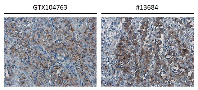
PD-L1 antibody detects PD-L1 protein at cell membrane in human ovarian carcinoma by immunohistochemical analysis. Antibodies: PD-L1 antibody diluted at 1:1000, and competitor's antibody diluted at 1:50.Application
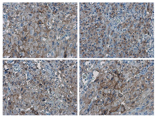
PD-L1 antibody detects PD-L1 proteinat cell membrane in human ovarian carcinoma by immunohistochemical analysis. Sample: Paraffin-embedded human ovarian carcinoma. PD-L1 antibody diluted at 1:1000.| Product Name | PD-L1 antibody |
|---|---|
| Antibody Type | Primary Antibodies |
| Product description | Rabbit Polyclonal antibody to PD-L1 (CD274 molecule)Involved in the costimulatory signal, essential for T-cell proliferation and production of IL10 and IFNG, in an IL2-dependent and a PDCD1-independent manner. Interaction with PDCD1 inhibits T-cell proliferation and cytokine production. |
| Immunogen | Carrier-protein conjugated synthetic peptide encompassing a sequence within the C-terminus region of human PD-L1. The exact sequence is proprietary. |
| Clonality | Polyclonal |
|---|---|
| Isotype | IgG |
| Host Species | Rabbit |
| Tested Applications | IHC-P,WB |
| *Optimal dilutions/concentrations should be determined by the researcher. | |
| Species Reactivity | Human |
| Concentration | 1mg/ml |
| Purification | Affinity purified |
| Gene Synonyms | B7-H antibody, B7H1 antibody, MGC142294 antibody, MGC142296 antibody, PD-L1 antibody, PDCD1L1 antibody, PDCD1LG1 antibody, PDL1 antibody, CD274 antibody, B7 homolog 1 antibody, programmed death ligand 1 antibody, programmed cell death 1 ligand 1 |
|---|---|
| Molecular Weight(MW) | 33 kDa (note) (2) |
| Cellular Localization | Isoform 1: Cell membrane; Single-pass type I membrane protein , Isoform 2: Endomembrane system; Single-pass type I membrane protein |
Application
Various whole cell extracts (30 µg) were separated by 10% SDS-PAGE, and the membrane was blotted with PD-L1 antibody diluted at 1:2000.
Application
Untreated (–) and treated (+) A431 whole cell extracts (30 µg) were separated by 10% SDS-PAGE, and the membranes were blotted with PD-L1 antibody diluted at 1:6000 and competitor's antibody (CST#13684) diluted at 1:500. The HRP-conjugated anti-rabbit IgG antibody was used to detect the primary antibody.
Application
Untreated (–) and treated (+) A549 whole cell extracts (30 µg) were separated by 10% SDS-PAGE, and the membranes were blotted with PD-L1 antibody diluted at 1:3000 and competitor's antibody (CST#13684) diluted at 1:500. The HRP-conjugated anti-rabbit IgG antibody was used to detect the primary antibody.
Application
Various whole cell extracts were separated by 10% SDS-PAGE, and the membranes were blotted with PD-L1 antibody diluted at 1:3000 and with DDDDK tag antibody diluted at 1:3000 to detect DDDDK-tagged PD-L2. The HRP-conjugated anti-rabbit IgG antibody was used to detect the primary antibody.
Application
Non-transfected (–) and transfected (+) A431 whole cell extracts (30 µg) were separated by 10% SDS-PAGE, and the membrane was blotted with PD-L1 antibody diluted at 1:5000. The HRP-conjugated anti-rabbit IgG antibody was used to detect the primary antibody.
Application
Untreated (–) and treated (+) MDA-MB-231 whole cell extracts (30 µg) were separated by 10% SDS-PAGE, and the membrane was blotted with PD-L1 antibody diluted at 1:5000.
Application
PD-L1 antibody detects PD-L1 protein at cell membrane in PD-L1 protein-expressing cell lines by immunohistochemical analysis. Antibodies: PD-L1 antibody diluted at 1:1000, and competitor's antibody diluted at 1:50. Samples: Negative (-), low positive (+), intermediate positive (++) and strong positive (+++) cell line cores assessed using Quantitative Digital Pathology.
Application
PD-L1 antibody detects PD-L1 protein at cell membrane in human ovarian carcinoma by immunohistochemical analysis. Antibodies: PD-L1 antibody diluted at 1:1000, and competitor's antibody diluted at 1:50.
Application
PD-L1 antibody detects PD-L1 proteinat cell membrane in human ovarian carcinoma by immunohistochemical analysis. Sample: Paraffin-embedded human ovarian carcinoma. PD-L1 antibody diluted at 1:1000.| Application Notes | *Optimal dilutions/concentrations should be determined by the researcher. |
|---|
| Form | Liquid |
|---|---|
| Storage Instructions | Keep as concentrated solution. Aliquot and store at -20ºC or below. Avoid multiple freeze-thaw cycles. |
| Storage Buffer | 1XPBS, 20% Glycerol (pH7). 0.025% ProClin 300 was added as a preservative. |


