Application
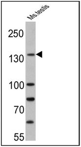
WB analysis of mouse testis lysate (25 ug) using PER1 antibody at a dilution of 1:500.Application
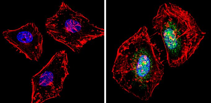
ICC/IF analysis of HeLa cells was performed in the presence (right) or absence (left) of PER1 antibody at a dilution of 1:100 (green). F-actin (red) was stained with a flourescent red phalloidin and nuclei (blue) were stained with Hoechst or DAPI. Images were taken at a magnification of 60x.Application
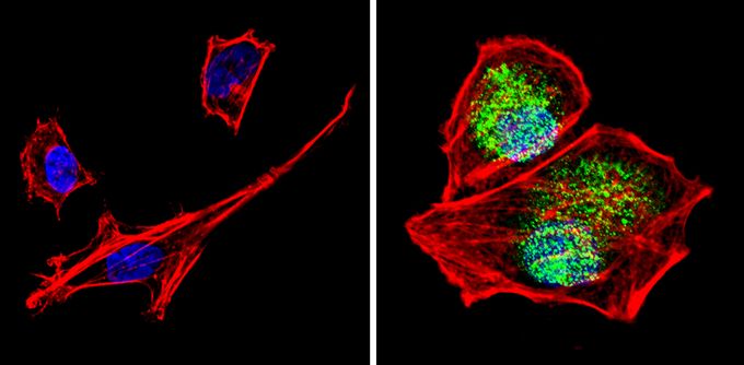
ICC/IF analysis of SH-SY5Y cells was performed in the presence (right) or absence (left) of PER1 antibody at a dilution of 1:100 (green). F-actin (red) was stained with a flourescent red phalloidin and nuclei (blue) were stained with Hoechst or DAPI. Images were taken at a magnification of 60x.Application
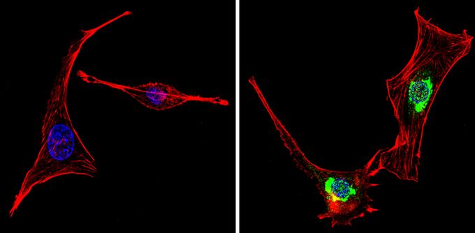
ICC/IF analysis of NIH-3T3 cells was performed in the presence (right) or absence (left) of PER1 antibody at a dilution of 1:100 (green). F-actin (red) was stained with a flourescent red phalloidin and nuclei (blue) were stained with Hoechst or DAPI. Images were taken at a magnification of 60x.Application
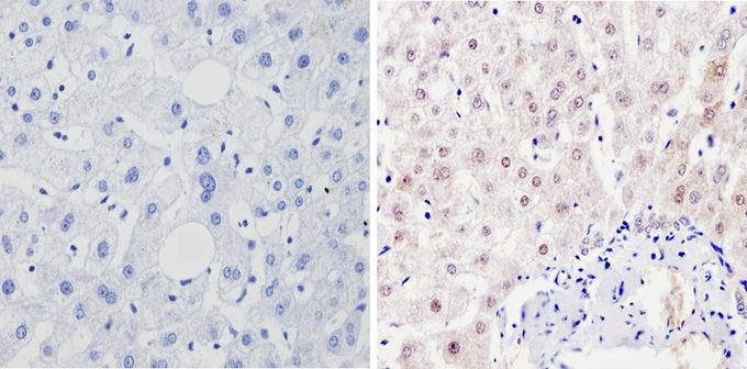
IHC-P analysis of human liver tissue was performed in the presence (right) or absence (left) of PER1 antibody at a dilution of 1:100.Application
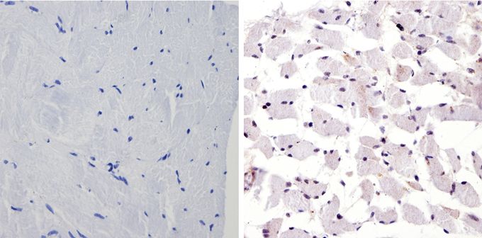
IHC-P analysis of human skeletal muscle was performed in the presence (right) or absence (left) of PER1 antibody at a dilution of 1:100.Application
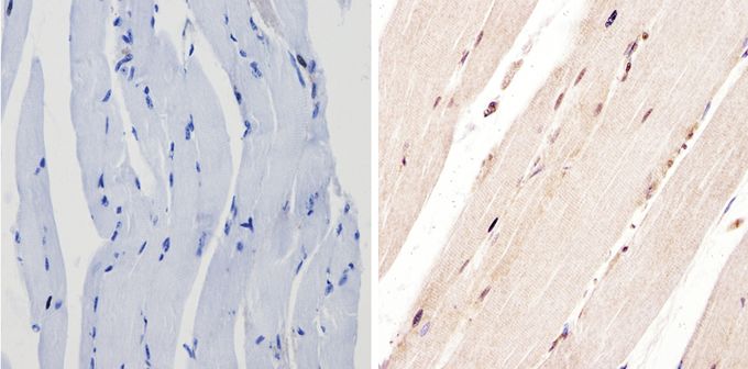
IHC-P analysis of mouse skeletal muscle was performed in the presence (right) or absence (left) of PER1 antibody at a dilution of 1:100.| Product Name | PER1 antibody |
|---|---|
| Antibody Type | Primary Antibodies |
| Product description | Rabbit polyclonal antibody to PER1This gene is a member of the Period family of genes and is expressed in a circadian pattern in the suprachiasmatic nucleus, the primary circadian pacemaker in the mammalian brain. Genes in this family encode components of the circadian rhythms of locomotor activity, metabolism, and behavior. Circadian expression in the suprachiasmatic nucleus continues in constant darkness, and a shift in the light/dark cycle evokes a proportional shift of gene expression in the suprachiasmatic nucleus. The specific function of this gene is not yet known. Alternative splicing has been observed in this gene; however, these variants have not been fully described. [provided by RefSeq, Jul 2008] |
| Immunogen | Synthetic Peptide: P(39) S L A D D T D A N S N G(51) C |
| Clonality | Polyclonal |
|---|---|
| Isotype | IgG |
| Host Species | Rabbit |
| Tested Applications | ICC/IFIHC-FIHC-PIPWB |
| Recommended Starting Dilutions: For ICC/IF: Use at a dilution of 1:10-1:200. For IHC-P: Use at a dilution of 1:50-1:500. For IHC-Fr: Use at a dilution of 1:50-1:500. For WB: Use at a dilution of 1:100 - 1:1000. Optimal dilution should be determined by the end user.: |
|
| Species Reactivity | HumanMouseDrosophilaRatHamster |
| Concentration | 1mg/ml |
| Purification | Affinity purified |
| Gene Synonyms | RIGUI PER period circadian clock 1 hPER |
|---|---|
| Target | PER1 |
Application
WB analysis of mouse testis lysate (25 ug) using PER1 antibody at a dilution of 1:500.
Application
ICC/IF analysis of HeLa cells was performed in the presence (right) or absence (left) of PER1 antibody at a dilution of 1:100 (green). F-actin (red) was stained with a flourescent red phalloidin and nuclei (blue) were stained with Hoechst or DAPI. Images were taken at a magnification of 60x.
Application
ICC/IF analysis of SH-SY5Y cells was performed in the presence (right) or absence (left) of PER1 antibody at a dilution of 1:100 (green). F-actin (red) was stained with a flourescent red phalloidin and nuclei (blue) were stained with Hoechst or DAPI. Images were taken at a magnification of 60x.
Application
ICC/IF analysis of NIH-3T3 cells was performed in the presence (right) or absence (left) of PER1 antibody at a dilution of 1:100 (green). F-actin (red) was stained with a flourescent red phalloidin and nuclei (blue) were stained with Hoechst or DAPI. Images were taken at a magnification of 60x.
Application
IHC-P analysis of human liver tissue was performed in the presence (right) or absence (left) of PER1 antibody at a dilution of 1:100.
Application
IHC-P analysis of human skeletal muscle was performed in the presence (right) or absence (left) of PER1 antibody at a dilution of 1:100.
Application
IHC-P analysis of mouse skeletal muscle was performed in the presence (right) or absence (left) of PER1 antibody at a dilution of 1:100.| Application Notes | Recommended Starting Dilutions: For ICC/IF: Use at a dilution of 1:10-1:200. For IHC-P: Use at a dilution of 1:50-1:500. For IHC-Fr: Use at a dilution of 1:50-1:500. For WB: Use at a dilution of 1:100 - 1:1000. Optimal dilution should be determined by the end user.: |
|---|
| Form | Liquid |
|---|---|
| Storage Instructions | -20° C, Avoid Freeze/Thaw Cycles. |
| Storage Buffer | PBS containing 1 mg/ml BSA, 0.05% sodium azide. |


