Application
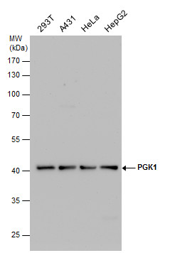
PGK1 antibody detects PGK1 protein by western blot analysis. Various whole cell extracts (30 µg) were separated by 10% SDS-PAGE, and the membrane was blotted with PGK1 antibody diluted by 1:1000. The HRP-conjugated anti-rabbit IgG antibody was used to detect the primary antibody.Application
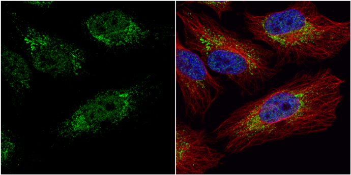
PGK1 antibody detects PGK1 protein at cytoplasm and nucleus by immunofluorescent analysis. Sample: HeLa cells were fixed in 4% paraformaldehyde at RT for 15 min. Green: PGK1 protein stained by PGK1 antibody diluted at 1:20. Red: alpha Tubulin, a cytoskeleton marker, stained by alpha Tubulin antibody [B-5-1-2] diluted at 1:10000. Blue: Hoechst 33342 staining.Application
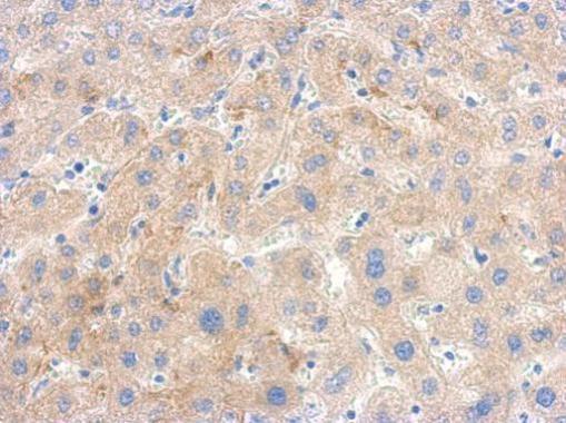
Immunohistochemical analysis of paraffin-embedded human hepatoma, using PGK1 antibody antibody at 1:500 dilution.Application
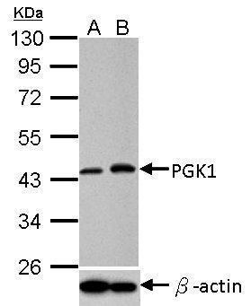
PGK1 antibody detects PGK1 protein in 1% O2-treated samples by western blot analysis. Upper panel: PGK1 antibody Lower panel: beta Actin antibody A. 30 µg HeLa whole cell lysate/extract (untreated) B. 30 µg HeLa whole cell lysate/extract (1% O2 treatment for 24hr) 10% SDS PAGE PGK1 antibody dilution: 1:1000 beta Actin antibody dilution: 1:10000 The HRP-conjugated anti-rabbit IgG antibody was used to detect the primary antibody.Application
Sample (50 µg of whole cell lysate) A: Mouse brain 10% SDS PAGE diluted at 1:1000 The HRP-conjugated anti-rabbit IgG antibody was used to detect the primary antibody.Application
PGK1 antibody detects PGK1 protein by western blot analysis. A. 50 µg Rat liver lysate/extract 10% SDS-PAGE PGK1 antibody dilution: 1:1000 The HRP-conjugated anti-rabbit IgG antibody was used to detect the primary antibody.Application
Immunofluorescence analysis of methanol-fixed HeLa, using PGK1 antibody at 1:50 dilution.| Product Name | PGK1 antibody |
|---|---|
| Antibody Type | Primary Antibodies |
| Product description | Rabbit Polyclonal antibody to PGK1 (phosphoglycerate kinase 1)The protein encoded by this gene is a glycolytic enzyme that catalyzes the conversion of 1,3-diphosphoglycerate to 3-phosphoglycerate. The encoded protein may also act as a cofactor for polymerase alpha. This gene lies on the X-chromosome, while a related pseudogene also has been found on the X-chromosome and another on chromosome 19. [provided by RefSeq] |
| Immunogen | Recombinant protein encompassing a sequence within the center region of human PGK1. The exact sequence is proprietary. |
| Clonality | Polyclonal |
|---|---|
| Isotype | IgG |
| Host Species | Rabbit |
| Tested Applications | ELISAICC/IFIHC-PWB |
| *Optimal dilutions/concentrations should be determined by the researcher.
: |
|
| Species Reactivity | HumanMouseRatYeast |
| Predicted Reactivity | Zebrafish, Xenopus laevis, Pig, Chicken, Sheep, Rhesus Monkey, Chimpanzee, BovineZebrafish (87%), Xenopus laevis (84%), Pig (96%), Chicken (88%), Sheep (96%), Rhesus Monkey (100%), Chimpanzee (100%), Bovine (96%) |
| Concentration | 1mg/ml |
| Purification | Affinity purified |
| Gene Synonyms | MGC117307 antibody MGC142128 antibody MGC8947 antibody MIG10 antibody PGKA antibody PGK1 antibody phosphoglycerate kinase 1 antibody cell migration-inducing gene 10 protein antibody PRP 2 antibody primer recognition protein 2 antibody |
|---|---|
| Molecular Weight(MW) | 45 kDa (note) (2) |
| Cellular Localization | Cytoplasm |
Application
PGK1 antibody detects PGK1 protein by western blot analysis. Various whole cell extracts (30 µg) were separated by 10% SDS-PAGE, and the membrane was blotted with PGK1 antibody diluted by 1:1000. The HRP-conjugated anti-rabbit IgG antibody was used to detect the primary antibody.
Application
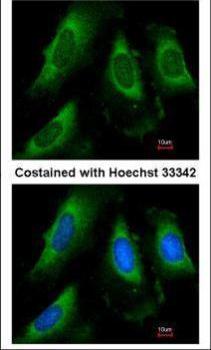
PGK1 antibody detects PGK1 protein at cytoplasm and nucleus by immunofluorescent analysis. Sample: HeLa cells were fixed in 4% paraformaldehyde at RT for 15 min. Green: PGK1 protein stained by PGK1 antibody diluted at 1:20. Red: alpha Tubulin, a cytoskeleton marker, stained by alpha Tubulin antibody [B-5-1-2] diluted at 1:10000. Blue: Hoechst 33342 staining.
Application
Immunohistochemical analysis of paraffin-embedded human hepatoma, using PGK1 antibody antibody at 1:500 dilution.
Application
PGK1 antibody detects PGK1 protein in 1% O2-treated samples by western blot analysis. Upper panel: PGK1 antibody Lower panel: beta Actin antibody A. 30 µg HeLa whole cell lysate/extract (untreated) B. 30 µg HeLa whole cell lysate/extract (1% O2 treatment for 24hr) 10% SDS PAGE PGK1 antibody dilution: 1:1000 beta Actin antibody dilution: 1:10000 The HRP-conjugated anti-rabbit IgG antibody was used to detect the primary antibody.
Application
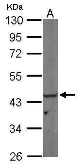
Sample (50 µg of whole cell lysate) A: Mouse brain 10% SDS PAGE diluted at 1:1000 The HRP-conjugated anti-rabbit IgG antibody was used to detect the primary antibody.
Application
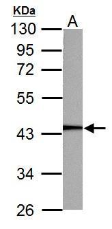
PGK1 antibody detects PGK1 protein by western blot analysis. A. 50 µg Rat liver lysate/extract 10% SDS-PAGE PGK1 antibody dilution: 1:1000 The HRP-conjugated anti-rabbit IgG antibody was used to detect the primary antibody.
Application
Immunofluorescence analysis of methanol-fixed HeLa, using PGK1 antibody at 1:50 dilution.| Positive Control | 293T , A431 , HeLa , HepG2 , mouse brain , Rat liver , *Yeast wild type 303 |
|---|---|
| Application Notes | *Optimal dilutions/concentrations should be determined by the researcher.
: |
| Form | Liquid |
|---|---|
| Storage Instructions | Keep as concentrated solution. Aliquot and store at -20ºC or below. Avoid multiple freeze-thaw cycles. |
| Storage Buffer | 1XPBS, 20% Glycerol (pH7). 0.025% ProClin 300 was added as a preservative. |


