Application
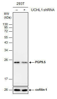
Non-transfected (–) and transfected (+) 293T whole cell extracts (30 µg) were separated by 12% SDS-PAGE, and the membrane was blotted with PGP9.5 antibody diluted at 1:3000.Application
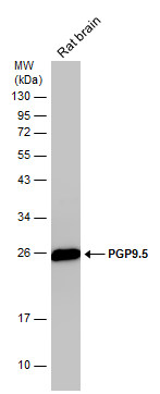
Rat tissue extract (50 µg) was separated by 12% SDS-PAGE, and the membrane was blotted with PGP9.5 antibody diluted at 1:10000.Application
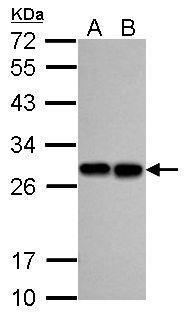
Sample (30 ug of whole cell lysate) A: A549 B: H1299 12% SDS PAGE diluted at 1:5000Application
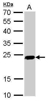
PGP9.5 antibody detects UCHL1 protein by Western blot analysis. A. 50 µg mouse brain lysate/extract 12 % SDS-PAGE PGP9.5 antibody dilution: 1:5000Application
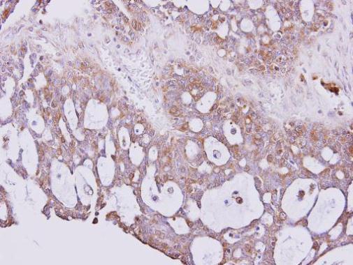
Immunohistochemical analysis of paraffin-embedded SEROUS OVCA xenograft, using PGP9.5 antibody at 1:300 dilution.Application
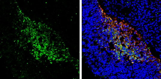
PGP9.5 antibody detects PGP9.5 protein expression by immunohistochemical analysis. Sample: Frozen sectioned E13.5 Rat brain. Green: PGP9.5 protein stained by PGP9.5 antibody diluted at 1:250. Red: beta Tubulin 3/ TUJ1, a mature neuron marker, stained by beta Tubulin 3/ TUJ1 antibody [GT11710] diluted at 1:500. Blue: Fluoroshield with DAPI .| Product Name | PGP9.5 antibody |
|---|---|
| Antibody Type | Primary Antibodies |
| Product description | Rabbit Polyclonal antibody to PGP9.5 (ubiquitin carboxyl-terminal esterase L1 (ubiquitin thiolesterase))UCHL1 is a member of a gene family whose products hydrolyze small C-terminal adducts of ubiquitin to generate the ubiquitin monomer. Expression of UCHL1 is highly specific to neurons and to cells of the diffuse neuroendocrine system and their tumors. It is present in all neurons (Doran et al., 1983 [PubMed 6343558]).[supplied by OMIM] |
| Immunogen | Carrier-protein conjugated synthetic peptide encompassing a sequence within the C-terminus region of human PGP9.5. The exact sequence is proprietary. |
| Clonality | Polyclonal |
|---|---|
| Isotype | IgG |
| Host Species | Rabbit |
| Tested Applications | IHC-FIHC-PWB |
| *Optimal dilutions/concentrations should be determined by the researcher.
: |
|
| Species Reactivity | HumanMouseRat |
| Predicted Reactivity | Xenopus laevis, Xenopus Tropicalis, Pig, BovineXenopus laevis (80%), Xenopus Tropicalis (81%), Pig (88%), Bovine (82%) |
| Concentration | 1mg/ml (Please refer to the vial label for the specific concentration) |
| Purification | Affinity purified |
| Gene Synonyms | PARK5 antibody PGP 9.5 antibody PGP9.5 antibody PGP95 antibody Uch-L1 antibody UCHL1 antibody ubiquitin thioesterase L1 antibody neuron cytoplasmic protein 9.5 antibody ubiquitin C-terminal hydrolase antibody ubiquitin carboxyl-terminal hydrolase |
|---|---|
| Molecular Weight(MW) | 25 kDa (note) |
| Cellular Localization | Cytoplasm |
Application
Non-transfected (–) and transfected (+) 293T whole cell extracts (30 µg) were separated by 12% SDS-PAGE, and the membrane was blotted with PGP9.5 antibody diluted at 1:3000.
Application
Rat tissue extract (50 µg) was separated by 12% SDS-PAGE, and the membrane was blotted with PGP9.5 antibody diluted at 1:10000.
Application
Sample (30 ug of whole cell lysate) A: A549 B: H1299 12% SDS PAGE diluted at 1:5000
Application
PGP9.5 antibody detects UCHL1 protein by Western blot analysis. A. 50 µg mouse brain lysate/extract 12 % SDS-PAGE PGP9.5 antibody dilution: 1:5000
Application
Immunohistochemical analysis of paraffin-embedded SEROUS OVCA xenograft, using PGP9.5 antibody at 1:300 dilution.
Application
PGP9.5 antibody detects PGP9.5 protein expression by immunohistochemical analysis. Sample: Frozen sectioned E13.5 Rat brain. Green: PGP9.5 protein stained by PGP9.5 antibody diluted at 1:250. Red: beta Tubulin 3/ TUJ1, a mature neuron marker, stained by beta Tubulin 3/ TUJ1 antibody [GT11710] diluted at 1:500. Blue: Fluoroshield with DAPI .| Positive Control | 293T , A549 , H1299 , mouse brain , rat brain |
|---|---|
| Application Notes | *Optimal dilutions/concentrations should be determined by the researcher.
: |
| Form | Liquid |
|---|---|
| Storage Instructions | Keep as concentrated solution. Aliquot and store at -20ºC or below. Avoid multiple freeze-thaw cycles. |
| Storage Buffer | 0.1M Tris, 0.1M Glycine, 10% Glycerol (pH7). 0.01% Thimerosal was added as a preservative. |


