Application
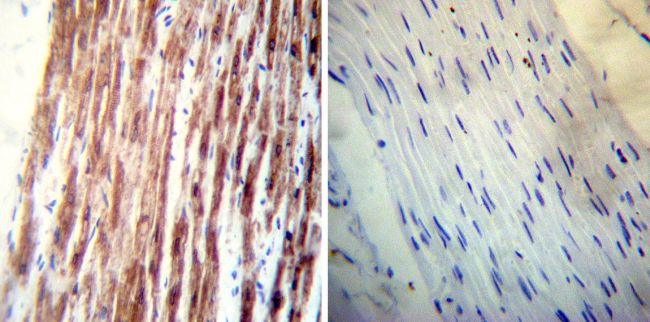
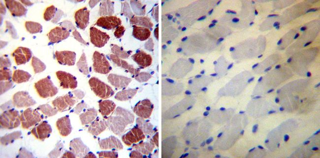
Immunohistochemistry was performed on normal deparaffinized Human skeletal muscle tissues. To expose target proteins, heat induced antigen retrieval was performed using 10mM sodium citrate (pH6.0) buffer, microwaved for 8-15 minutes. Following antigen retrieval tissues were blocked in 3% BSA-PBS for 30 minutes at room temperature. Tissues were then probed at a dilution of 1:200 with a mouse monoclonal antibody recognizing Phospholamban or without primary antibody (negative control) overnight at 4°C in a humidified chamber. Tissues were washed extensively with PBST and endogenous peroxidase activity was quenched with a peroxidase suppressor. Detection was performed using a biotin-conjugated secondary antibody and SA-HRP, followed by colorimetric detection using DAB. Tissues were counterstained with hematoxylin and prepped for mounting.Application
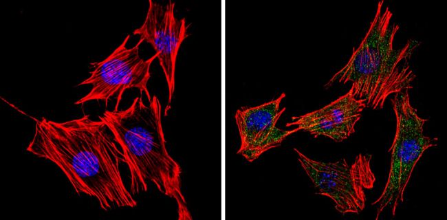
Immunofluorescent analysis of Phospholamban (green) showing staining in the cytoplasm of C2C12 cells (right) compared to a negative control without primary antibody (left). Formalin-fixed cells were permeabilized with 0.1% Triton X-100 in TBS for 5-10 minutes and blocked with 3% BSA-PBS for 30 minutes at room temperature. Cells were probed with a Phospholamban monoclonal antibody in 3% BSA-PBS at a dilution of 1:20 and incubated overnight at 4°C in a humidified chamber. Cells were washed with PBST and incubated with a DyLight-conjugated secondary antibody in PBS at room temperature in the dark. Actin was stained using Alexa Fluor 554 (red) and nuclei were stained with Hoechst or DAPI (blue). Images were taken at a magnification of 60x.Application
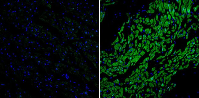
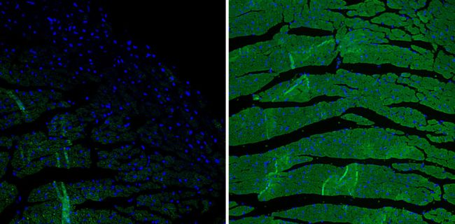
Immunofluorescent analysis of Phospholamban (green) showing staining in the cytoplasm of mouse heart tissue (right) compared to a negative control without primary antibody (left). Formalin-fixed tissue was permeabilized with 0.1% Triton X-100 in TBS for 5-10 minutes and blocked with 3% BSA-PBS for 30 minutes at room temperature. Tissue was probed with a Phospholamban monoclonal antibody in 3% BSA-PBS at a dilution of 1:100 and incubated overnight at 4°C in a humidified chamber. Tissue was washed with PBST and incubated with a DyLight-conjugated secondary antibody in PBS at room temperature in the dark. Nuclei were stained with Hoechst or DAPI (blue). Images were taken at a magnification of 60x.Application
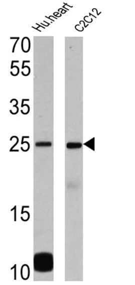
Western blot analysis of Phospholamban was performed by loading 25 ug of human heart (lane 1) and C2C12 (lane 2) onto an SDS polyacrylamide gel. Proteins were transferred to a PVDF membrane and blocked at 4°C overnight. The membrane was probed with a Phospholamban monoclonal antibody at a dilution of 1:1000 overnight at 4°C, washed in TBST, and probed with an HRP-conjugated secondary antibody for 1 hr at room temperature in the dark. Chemiluminescent detection was performed using Pierce ECL Plus Western Blotting Substrate. Results show a band at ~25kDa.Application
Immunofluorescent analysis of Phospholamban (green) showing staining in the cytoplasm of human heart tissue (right) compared to a negative control without primary antibody (left). Formalin-fixed tissue was permeabilized with 0.1% Triton X-100 in TBS for 5-10 minutes and blocked with 3% BSA-PBS for 30 minutes at room temperature. Tissue was probed with a Phospholamban monoclonal antibody in 3% BSA-PBS at a dilution of 1:20 and incubated overnight at 4°C in a humidified chamber. Tissue was washed with PBST and incubated with a DyLight-conjugated secondary antibody in PBS at room temperature in the dark. Nuclei were stained with Hoechst or DAPI (blue). Images were taken at a magnification of 60x.Application
Immunohistochemistry was performed on normal deparaffinized Human heart tissue tissues. To expose target proteins, heat induced antigen retrieval was performed using 10mM sodium citrate (pH6.0) buffer, microwaved for 8-15 minutes. Following antigen retrieval tissues were blocked in 3% BSA-PBS for 30 minutes at room temperature. Tissues were then probed at a dilution of 1:200 with a mouse monoclonal antibody recognizing Phospholamban or without primary antibody (negative control) overnight at 4°C in a humidified chamber. Tissues were washed extensively with PBST and endogenous peroxidase activity was quenched with a peroxidase suppressor. Detection was performed using a biotin-conjugated secondary antibody and SA-HRP, followed by colorimetric detection using DAB. Tissues were counterstained with hematoxylin and prepped for mounting.| Product Name | Phospholamban antibody |
|---|---|
| Antibody Type | Primary Antibodies |
| Product description | Mouse monoclonal antibody [2D12] to Phospholamban |
| Immunogen | Synthetic peptide corresponding to residues D(2) K V Q Y L T R S A I R R A S T I E M P Q Q A R(25) of canine PLB. |
| Clonality | Monoclonal |
|---|---|
| Isotype | IgG2a |
| Clone Number | 2D12 |
| Host Species | Mouse |
| Tested Applications | BlockingICC/IFIHCIHC-PIPWB |
| IHC-P: 1:200: |
|
| Species Reactivity | HumanMouseChickenDogPigRatGuinea PigRabbitSheep |
| Concentration | 1mg/ml (Please refer to the vial label for the specific concentration) |
| Gene Synonyms | phospholamban PLN |
|---|---|
| Target | Phospholamban |
Application
Immunohistochemistry was performed on normal deparaffinized Human skeletal muscle tissues. To expose target proteins, heat induced antigen retrieval was performed using 10mM sodium citrate (pH6.0) buffer, microwaved for 8-15 minutes. Following antigen retrieval tissues were blocked in 3% BSA-PBS for 30 minutes at room temperature. Tissues were then probed at a dilution of 1:200 with a mouse monoclonal antibody recognizing Phospholamban or without primary antibody (negative control) overnight at 4°C in a humidified chamber. Tissues were washed extensively with PBST and endogenous peroxidase activity was quenched with a peroxidase suppressor. Detection was performed using a biotin-conjugated secondary antibody and SA-HRP, followed by colorimetric detection using DAB. Tissues were counterstained with hematoxylin and prepped for mounting.
Application
Immunofluorescent analysis of Phospholamban (green) showing staining in the cytoplasm of C2C12 cells (right) compared to a negative control without primary antibody (left). Formalin-fixed cells were permeabilized with 0.1% Triton X-100 in TBS for 5-10 minutes and blocked with 3% BSA-PBS for 30 minutes at room temperature. Cells were probed with a Phospholamban monoclonal antibody in 3% BSA-PBS at a dilution of 1:20 and incubated overnight at 4°C in a humidified chamber. Cells were washed with PBST and incubated with a DyLight-conjugated secondary antibody in PBS at room temperature in the dark. Actin was stained using Alexa Fluor 554 (red) and nuclei were stained with Hoechst or DAPI (blue). Images were taken at a magnification of 60x.
Application
Immunofluorescent analysis of Phospholamban (green) showing staining in the cytoplasm of mouse heart tissue (right) compared to a negative control without primary antibody (left). Formalin-fixed tissue was permeabilized with 0.1% Triton X-100 in TBS for 5-10 minutes and blocked with 3% BSA-PBS for 30 minutes at room temperature. Tissue was probed with a Phospholamban monoclonal antibody in 3% BSA-PBS at a dilution of 1:100 and incubated overnight at 4°C in a humidified chamber. Tissue was washed with PBST and incubated with a DyLight-conjugated secondary antibody in PBS at room temperature in the dark. Nuclei were stained with Hoechst or DAPI (blue). Images were taken at a magnification of 60x.
Application
Western blot analysis of Phospholamban was performed by loading 25 ug of human heart (lane 1) and C2C12 (lane 2) onto an SDS polyacrylamide gel. Proteins were transferred to a PVDF membrane and blocked at 4°C overnight. The membrane was probed with a Phospholamban monoclonal antibody at a dilution of 1:1000 overnight at 4°C, washed in TBST, and probed with an HRP-conjugated secondary antibody for 1 hr at room temperature in the dark. Chemiluminescent detection was performed using Pierce ECL Plus Western Blotting Substrate. Results show a band at ~25kDa.
Application
Immunofluorescent analysis of Phospholamban (green) showing staining in the cytoplasm of human heart tissue (right) compared to a negative control without primary antibody (left). Formalin-fixed tissue was permeabilized with 0.1% Triton X-100 in TBS for 5-10 minutes and blocked with 3% BSA-PBS for 30 minutes at room temperature. Tissue was probed with a Phospholamban monoclonal antibody in 3% BSA-PBS at a dilution of 1:20 and incubated overnight at 4°C in a humidified chamber. Tissue was washed with PBST and incubated with a DyLight-conjugated secondary antibody in PBS at room temperature in the dark. Nuclei were stained with Hoechst or DAPI (blue). Images were taken at a magnification of 60x.
Application
Immunohistochemistry was performed on normal deparaffinized Human heart tissue tissues. To expose target proteins, heat induced antigen retrieval was performed using 10mM sodium citrate (pH6.0) buffer, microwaved for 8-15 minutes. Following antigen retrieval tissues were blocked in 3% BSA-PBS for 30 minutes at room temperature. Tissues were then probed at a dilution of 1:200 with a mouse monoclonal antibody recognizing Phospholamban or without primary antibody (negative control) overnight at 4°C in a humidified chamber. Tissues were washed extensively with PBST and endogenous peroxidase activity was quenched with a peroxidase suppressor. Detection was performed using a biotin-conjugated secondary antibody and SA-HRP, followed by colorimetric detection using DAB. Tissues were counterstained with hematoxylin and prepped for mounting.| Application Notes | IHC-P: 1:200: |
|---|
| Form | Liquid |
|---|---|
| Storage Instructions | -20° C, Avoid Freeze/Thaw Cycles |
| Storage Buffer | Ascites containing 0.05 % sodium azide |


