Application
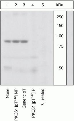
Lysates prepared from K562 cells stimulated with PMA were resolved by SDS-PAGE on a 10% polyacrylamide gel and transferred to PVDF. Membranes were either left untreated (1-4) or treated with lambda phosphatase (5), blocked with a 5% BSA-TBST buffer for one hour at room temperature, and incubated with PKCbetaI [pT642] antibody for two hours at room temperature in a 3% BSA-TBST buffer, following prior incubation with: no peptide (1, 5), the non-phosphopeptide corresponding to the immunogen (2), a generic phosphothreonine-containing peptide (3), or, the phosphopeptide immunogen (4). After washing, membranes were incubated with goat F(ab)2 anti-rabbit IgG HRP conjugate and bands were detected. The data show that only the peptide corresponding to PKCbetaI [pT642] blocks the antibody signal. The data also show that phosphatase stripping eliminates the signal, verifying that the antibody is phospho-specific.| Product Name | PKC beta 1 (phospho Thr642) antibody |
|---|---|
| Antibody Type | Primary Antibodies |
| Product description | Rabbit polyclonal to PKC beta 1 ( phospho T642 )Protein Kinase C beta 1 (PKC beta 1) is an 80 kDa member of the conventional group (cPKCs: sensitive to diacylglycerol, phosphotidylserine and phorbol esters) of the PKC family of serine/threonine kinases that are involved in a wide range of physiological processes including mitogenesis, cell survival, transcriptional regulation and tumor promotion. PKC beta 1 is phosphorylated on three sites, threonine 500 in the activation loop, threonine 642 in the turn loop and serine 661 in the hydrophobic loop. Autophosphorylation of PKC beta 1 on threonine 642 is crucial for its activation and catalytic activity. Threonine 642 mediates PKC beta 1 ability to activate c-Fos in response to phorbol esters. |
| Immunogen | Synthetic phosphopeptide derived from a region of human PKC beta 1 that contains threonine 642. |
| Modification | p-Thr642 |
| Clonality | Polyclonal |
|---|---|
| Isotype | IgG |
| Host Species | Rabbit |
| Tested Applications | WB |
| WB: Use at a dilution of 1/1000. Detects a band of approximately 80 kDa. Optimal dilutions/concentrations should be determined by the end user. | |
| Species Reactivity | HumanRat |
| Concentration | 1mg/ml |
| Purification | Affinity purified |
| Gene Synonyms | MGC41878, PKC-beta, PKCB, PRKCB1, PRKCB2, PKC beta, PKCbeta, PKC beta2 (phospho T642), PKC beta2 (phospho Thr642), PKC beta2 (pT642), PKC beta2 (pThr642), PKC beta2 phospho T642, PKC beta2 phospho Thr642, Phospho PKC beta2 (T642), Phospho PKC beta2 (Thr64 |
|---|---|
| Target | PKC beta 1 |
| Tissue Specificity | This antibody does not react with PKC beta 2 [pT641], alpha [pT638], nu [pT655], epsilon [pT710], iota [pT555], eta [pT655], or zeta [pT560] as determined by peptide competition experiments. |
| Cellular Localization | Cytoplasmic and Plasma membrane |
Application
Lysates prepared from K562 cells stimulated with PMA were resolved by SDS-PAGE on a 10% polyacrylamide gel and transferred to PVDF. Membranes were either left untreated (1-4) or treated with lambda phosphatase (5), blocked with a 5% BSA-TBST buffer for one hour at room temperature, and incubated with PKCbetaI [pT642] antibody for two hours at room temperature in a 3% BSA-TBST buffer, following prior incubation with: no peptide (1, 5), the non-phosphopeptide corresponding to the immunogen (2), a generic phosphothreonine-containing peptide (3), or, the phosphopeptide immunogen (4). After washing, membranes were incubated with goat F(ab)2 anti-rabbit IgG HRP conjugate and bands were detected. The data show that only the peptide corresponding to PKCbetaI [pT642] blocks the antibody signal. The data also show that phosphatase stripping eliminates the signal, verifying that the antibody is phospho-specific.| Application Notes | WB: Use at a dilution of 1/1000. Detects a band of approximately 80 kDa. Optimal dilutions/concentrations should be determined by the end user. |
|---|
| Form | Liquid |
|---|---|
| Storage Instructions | Keep as concentrated solution. Store at 4ºC short term. For extended storage aliquot and store at -20ºC or below. Avoid freeze-thaw cycles. |
| Storage Buffer | PBS pH 7.3 containing 1mg/ml BSA, 50% glycerol and 0.05% sodium azide |


