Application
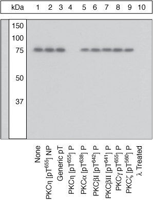
Lysates prepared from Jurkat cells stimulated with PMA were resolved by SDS-PAGE on a 10% polyacrylamide gel and transferred to PVDF. Membranes were either left untreated (1-9) or treated with lambda phosphatase (10), blocked with a 5% BSA-TBST buffer overnight at 4°C, and incubated with 0.35 µg/ml PKCeta [pT655] antibody for two hours at room temperature in a 3% BSA-TBST buffer, following prior incubation with: no peptide (1, 10), the non-phosphopeptide corresponding to the immunogen (2), a generic phosphothreonine containing peptide (3), the phosphopeptide immunogen (4), or, the phosphopeptide corresponding to the immunogen from other PKC isoforms (5-9). After washing, membranes were incubated with goat F(ab)2 anti-rabbit IgG HRP-conjugate and bands were detected. The data show that the peptide corresponding to PKCeta [pT655] blocks the antibody signal. The antibody signal was not blocked by the peptides corresponding to PKC isoforms alpha [pT638], betaI [pT642], betaI [pT641], gamma [pT655] and zeta [pT560], thereby demonstrating the specificity of the antibody. The data also show that phosphatase stripping eliminates the signal, verifying that the antibody is phospho-specific.| Product Name | PKC eta (phospho Thr655) antibody |
|---|---|
| Antibody Type | Primary Antibodies |
| Product description | Rabbit polyclonal to PKC eta ( phospho T655 )Protein Kinase C eta (PKC eta) is an 80 kDa member of the novel group (nPKCs: sensitive to diacylglycerol, phosphatidylserine and phorbol esters) of the PKC family of serine/threonine kinases that are involved in a wide range of physiological processes including mitogenesis, cell survival, transcriptional regulation and tumor promotion. PKC eta has been shown to be involved in the cell cycle as ectopic expression results in elevated expression of cyclins D and E, and its association with the cyclinE/cdk2/p21 complex inhibits cdk2 kinase activity, leading to G1 arrest. PKC eta is also involved in mediating FGF and PDGF ability to stimulate IL-6 production, and PKC eta modulation of ERK and JNK signaling pathways. The activation loop threonine (threonine 512 in PKC eta) of novel PKCs is phosphorylated by phosphoinositide dependent kinase-1 (PDK1) and is necessary for the autophosphorylation of threonine 655 in the carboxy-terminus of PKC eta, a step that may play a role in mediating PKC eta signaling events. |
| Immunogen | Synthetic phosphopeptide derived from a region of human PKC eta that contains threonine 655. |
| Modification | p-Thr655 |
| Clonality | Polyclonal |
|---|---|
| Isotype | IgG |
| Host Species | Rabbit |
| Tested Applications | WB |
| WB: Use at a concentration of 0.35 - 1.0 μg/ml. Detects a band of approximately 80 kDa. Optimal dilutions/concentrations should be determined by the end user. | |
| Species Reactivity | HumanMouseRat |
| Concentration | 1mg/ml |
| Purification | Affinity purified |
| Gene Synonyms | MGC26269, MGC5363, PKC-L, PKCL, PRKCL, nPKC-eta, PKC L, nPKC eta, nPKCeta, PKC eta (phospho T655), PKC eta (phospho Thr655), PKC eta (pT655), PKC eta (pThr655), PKC eta phospho T655, PKC eta phospho Thr655, Phospho PKC eta (T655), Phospho PKC eta (Thr655) |
|---|---|
| Target | PKC eta |
| Tissue Specificity | This antibody does not cross-react with any other PKC isoforms tested. |
Application
Lysates prepared from Jurkat cells stimulated with PMA were resolved by SDS-PAGE on a 10% polyacrylamide gel and transferred to PVDF. Membranes were either left untreated (1-9) or treated with lambda phosphatase (10), blocked with a 5% BSA-TBST buffer overnight at 4°C, and incubated with 0.35 µg/ml PKCeta [pT655] antibody for two hours at room temperature in a 3% BSA-TBST buffer, following prior incubation with: no peptide (1, 10), the non-phosphopeptide corresponding to the immunogen (2), a generic phosphothreonine containing peptide (3), the phosphopeptide immunogen (4), or, the phosphopeptide corresponding to the immunogen from other PKC isoforms (5-9). After washing, membranes were incubated with goat F(ab)2 anti-rabbit IgG HRP-conjugate and bands were detected. The data show that the peptide corresponding to PKCeta [pT655] blocks the antibody signal. The antibody signal was not blocked by the peptides corresponding to PKC isoforms alpha [pT638], betaI [pT642], betaI [pT641], gamma [pT655] and zeta [pT560], thereby demonstrating the specificity of the antibody. The data also show that phosphatase stripping eliminates the signal, verifying that the antibody is phospho-specific.| Application Notes | WB: Use at a concentration of 0.35 - 1.0 μg/ml. Detects a band of approximately 80 kDa. Optimal dilutions/concentrations should be determined by the end user. |
|---|
| Form | Liquid |
|---|---|
| Storage Instructions | Keep as concentrated solution. Store at 4ºC short term. For extended storage aliquot and store at -20ºC or below. Avoid freeze-thaw cycles. |
| Storage Buffer | Preservative: 0.05% Sodium Azide; Constituents: PBS, 1mg/ml BSA. pH 7.3 |


