Application
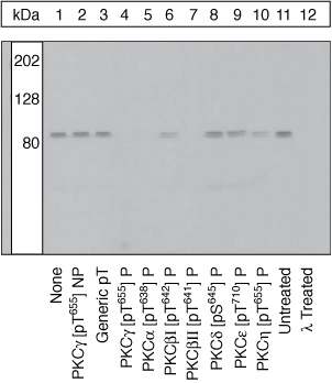
Lysates prepared from HeLa cells stimulated with PMA were resolved by SDS-PAGE on a 10% polyacrylamide gel and transferred to PVDF. Membranes were either left untreated (1-11) or treated with lambda phosphatase (12), blocked with a 3% low-fat milk-TBST buffer overnight at 4°C, and incubated with 0.50 µg/ml PKCgamma [pT655] antibody for two hours at room temperature in a 3% BSA-TBST buffer, following prior incubation with: no peptide (1, 11, 12), the non-phosphopeptide corresponding to the immunogen (2), a generic phosphothreonine containing peptide (3), the phosphopeptide immunogen (4), or, the phosphopeptide corresponding to the immunogen from other PKC isoforms (5-10). After washing, membranes were incubated with goat F(ab)2 anti-rabbit IgG alkaline phosphatase and signals were detected. The data show that the peptide corresponding to PKCgamma [pT655] blocks the antibody signal while the peptides corresponding to PKC isoforms betaI [pT642], delta [pS645], epsilon [pT710] and eta [pT655] do not completely block the antibody signal, thereby demonstrating the specificity of the antibody. However, peptides corresponding to PKC isoforms alpha [pT638] and betaII [pT641] do block the antibody signal, indicating that the PKCgamma [pT655] antibody may cross-react with their respective sites. The data also show that phosphatase stripping eliminates the signal, verifying that the antibody is phospho-specific.| Product Name | PKC gamma (phospho Thr655) antibody |
|---|---|
| Antibody Type | Primary Antibodies |
| Product description | Rabbit polyclonal to PKC gamma ( phospho T655 )PKC gamma is an 80 kDa member of the conventional group (cPKCs: sensitive to calcium, diacylglycerol and phorbol esters) of the PKC family of serine/threonine family kinases that are involved in a wide range of physiological processes including mitogenesis, cell survival and transcriptional regulation. PKC gamma plays a key role in neuronal signal transduction and is translocated from the nucleus to the cytoplasm upon activation by phorbol ester, where in epithelial cells it has been implicated in regulating intracellular communication. The activation loop threonine (threonine 514 in PKC gamma) of conventional PKCs is phosphorylated by phosphoinositide-dependent kinase-1 (PDK1), which is necessary for their autophosphorylation, a critical step in the generation of a catalytically mature enzyme. Threonine 655 is an autophosphorylation site in the carboxy-terminus of PKC gamma. |
| Immunogen | Synthetic phosphopeptide derived from a region of human PKC gamma that contains threonine 655. |
| Modification | p-Thr655 |
| Clonality | Polyclonal |
|---|---|
| Isotype | IgG |
| Host Species | Rabbit |
| Tested Applications | WB |
| WB: Use at a concentration of 0.5 - 1.0 μg/ml. Detects a band of approximately 80 kDa. Optimal dilutions/concentrations should be determined by the end user. | |
| Species Reactivity | HumanMouseRat |
| Concentration | 0.5mg/ml |
| Purification | Affinity purified |
| Gene Synonyms | MGC57564, PKC-gamma, PKCC, PKCG, SCA14, PKC gamma, PKCgamma, PKC gamma (phospho T655), PKC gamma (phospho Thr655), PKC gamma (pT655), PKC gamma (pThr655), PKC gamma phospho T655, PKC gamma phospho Thr655, Phospho PKC gamma (T655), Phospho PKC gamma (Thr65 |
|---|---|
| Target | PKC gamma |
| Tissue Specificity | PKC alpha (69%) and beta 2 (62%) may cross-react in cells expressing high levels of these proteins. |
| Cellular Localization | Cytoplasmic |
Application
Lysates prepared from HeLa cells stimulated with PMA were resolved by SDS-PAGE on a 10% polyacrylamide gel and transferred to PVDF. Membranes were either left untreated (1-11) or treated with lambda phosphatase (12), blocked with a 3% low-fat milk-TBST buffer overnight at 4°C, and incubated with 0.50 µg/ml PKCgamma [pT655] antibody for two hours at room temperature in a 3% BSA-TBST buffer, following prior incubation with: no peptide (1, 11, 12), the non-phosphopeptide corresponding to the immunogen (2), a generic phosphothreonine containing peptide (3), the phosphopeptide immunogen (4), or, the phosphopeptide corresponding to the immunogen from other PKC isoforms (5-10). After washing, membranes were incubated with goat F(ab)2 anti-rabbit IgG alkaline phosphatase and signals were detected. The data show that the peptide corresponding to PKCgamma [pT655] blocks the antibody signal while the peptides corresponding to PKC isoforms betaI [pT642], delta [pS645], epsilon [pT710] and eta [pT655] do not completely block the antibody signal, thereby demonstrating the specificity of the antibody. However, peptides corresponding to PKC isoforms alpha [pT638] and betaII [pT641] do block the antibody signal, indicating that the PKCgamma [pT655] antibody may cross-react with their respective sites. The data also show that phosphatase stripping eliminates the signal, verifying that the antibody is phospho-specific.| Application Notes | WB: Use at a concentration of 0.5 - 1.0 μg/ml. Detects a band of approximately 80 kDa. Optimal dilutions/concentrations should be determined by the end user. |
|---|
| Form | Liquid |
|---|---|
| Storage Instructions | Aliquot and store at -20°C or -80°C. Avoid repeated freeze / thaw cycles. |
| Storage Buffer | Preservative: 0.05% Sodium Azide; Constituents: PBS, 1mg/ml BSA. pH 7.3 |


