Application
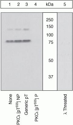
Western blot using PKC-iota [pT555]/PKC Lambda [pT563] Polyclonal Antibody, Rabbit . Lysates prepared from Jurkat cells stimulated with PMA were resolved by SDS-PAGE on a 10% polyacrylamide gel and transferred to PVDF. Membranes were either left untreated (1-4) or treated with lambda phosphatase (5), blocked with a 5% low-fat milk-TBST buffer for one hour at room temperature, and incubated with PKCiota [pT555] antibody for two hours at room temperature in a 3% low-fat milk-TBST buffer, following prior incubation with: no peptide (1), the non-phosphopeptide corresponding to the immunogen (2), a generic phosphothreonine-containing peptide (3), or, the phosphopeptide immunogen (4). After washing, membranes were incubated with goat F(ab’)2 anti-rabbit IgG HRP conjugate and bands were detected. The data show that the phosphopeptide corresponding to PKCiota [pT555] blocks the antibody signal. The peptide corresponding to PKCzeta [pT560] blocks the antibody signal and the peptides corresponding to PKC isoforms betaI [pT642] and gamma [pT655] partially block the antibody signal (data not shown), suggesting cross-reactivity of the antibody with these sites. The antibody signal was not blocked by the corresponding peptides of any other PKC isoforms. The data also show that phosphatase stripping eliminates the signal, verifying that the antibody is phospho-specific| Product Name | PKC iota + PKC lambda (phospho Thr555/Thr563) antibody |
|---|---|
| Antibody Type | Primary Antibodies |
| Product description | Rabbit polyclonal to PKC iota + PKC lambda ( phospho T555 + T563 )Protein Kinase C iota (PKC iota) is a 76 kDa member of the atypical group (cPKCs: sensitive to diacylglycerol, phosphotidylserine and phorbol esters) of the PKC family of serine/threonine kinases that are involved in a wide range of physiological processes including mitogenesis, cell survival, transcriptional regulation and tumor promotion. Phosphorylation of PKC iota plays a key role in protein trafficking. Phosphorylated PKC iota is implicated in high glucose-induced T type II transforming growth factor-beta activity, TNF-alpha-stimulated NF-kappaB activation and Bcr/Abl mediated anti-apoptotic effect. |
| Immunogen | Synthetic phosphopeptide derived from a region of human PKC iota that contains threonine 555. |
| Modification | p-Thr555/Thr563 |
| Clonality | Polyclonal |
|---|---|
| Isotype | IgG |
| Host Species | Rabbit |
| Tested Applications | ICC/IFWB |
| For WB: Use at a dilution of 1:1,000. Detects a band of approximately 76 kDa. Optimal dilutions/concentrations should be determined by the researcher. | |
| Species Reactivity | HumanMouse |
| Concentration | 1mg/ml |
| Purification | Affinity purified |
| Gene Synonyms | DXS1179E, MGC26534, PKCI, nPKC-iota, nPKC iota, nPKCiota, PKC iota + PKC lambda (phospho T555/T563), PKC iota + PKC lambda (phospho Thr555/Thr563), PKC iota + PKC lambda (pT555/pT563), PKC iota + PKC lambda (pThr555/pThr563), PKC iota + PKC lambda phospho |
|---|---|
| Target | PKC iota + PKC lambda |
| Tissue Specificity | This antibody reacts with PKC lambda immunoprecipitates, indicating cross-reactivity for PKC lambda [pT563]. PKC zeta [pT560] (83% homologous) has been shown to cross-react by peptide competition. Peptide competition also suggests that this antibody may partially cross-react with PKC beta 1 [pS642] (58% homologous) and PKC nu [pT655] (42% homologous). |
Application
Western blot using PKC-iota [pT555]/PKC Lambda [pT563] Polyclonal Antibody, Rabbit . Lysates prepared from Jurkat cells stimulated with PMA were resolved by SDS-PAGE on a 10% polyacrylamide gel and transferred to PVDF. Membranes were either left untreated (1-4) or treated with lambda phosphatase (5), blocked with a 5% low-fat milk-TBST buffer for one hour at room temperature, and incubated with PKCiota [pT555] antibody for two hours at room temperature in a 3% low-fat milk-TBST buffer, following prior incubation with: no peptide (1), the non-phosphopeptide corresponding to the immunogen (2), a generic phosphothreonine-containing peptide (3), or, the phosphopeptide immunogen (4). After washing, membranes were incubated with goat F(ab’)2 anti-rabbit IgG HRP conjugate and bands were detected. The data show that the phosphopeptide corresponding to PKCiota [pT555] blocks the antibody signal. The peptide corresponding to PKCzeta [pT560] blocks the antibody signal and the peptides corresponding to PKC isoforms betaI [pT642] and gamma [pT655] partially block the antibody signal (data not shown), suggesting cross-reactivity of the antibody with these sites. The antibody signal was not blocked by the corresponding peptides of any other PKC isoforms. The data also show that phosphatase stripping eliminates the signal, verifying that the antibody is phospho-specific| Application Notes | For WB: Use at a dilution of 1:1,000. Detects a band of approximately 76 kDa. Optimal dilutions/concentrations should be determined by the researcher. |
|---|
| Form | Liquid |
|---|---|
| Storage Instructions | Keep as concentrated solution. Store at 4°C short term. For extended storage aliquot and store at -20°C or below. Avoid freeze-thaw cycles. |
| Storage Buffer | Phosphate-buffered saline, pH 7.3, 50% glycerol contiaining 1.0 mg/ml BSA |


