Application
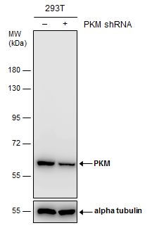
Non-transfected (–) and transfected (+) 293T whole cell extracts (30 µg) were separated by 7.5% SDS-PAGE, and the membrane was blotted with PKM antibody diluted at 1:5000. The HRP-conjugated anti-rabbit IgG antibody was used to detect the primary antibody.Application
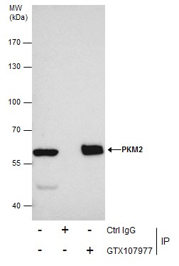
Immunoprecipitation of PKM2 protein from 293T whole cell extracts using 5 μg of Pyruvate Kinase (muscle) antibody . Western blot analysis was performed using Pyruvate Kinase (muscle) antibody . EasyBlot anti-Rabbit IgG was used as a secondary reagent.Application
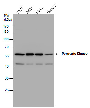
Pyruvate Kinase (muscle) antibody detects Pyruvate Kinase (muscle) protein by western blot analysis. Various whole cell extracts (30 µg) were separated by 10% SDS-PAGE, and the membrane was blotted with Pyruvate Kinase (muscle) antibody diluted by 1:1000. The HRP-conjugated anti-rabbit IgG antibody was used to detect the primary antibody.Application
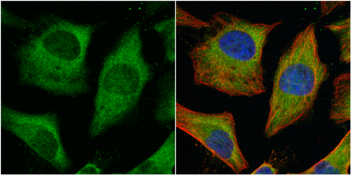
Pyruvate Kinase (muscle) antibody detects Pyruvate Kinase (muscle) protein at cytoplasm by immunofluorescent analysis. Sample: HeLa cells were fixed in 4% paraformaldehyde at RT for 15 min. Green: Pyruvate Kinase (muscle) protein stained by Pyruvate Kinase (muscle) antibody diluted at 1:500. Red: alpha Tubulin, a cytoskeleton marker, stained by alpha Tubulin antibody [GT114] diluted at 1:500. Blue: Hoechst 33342 staining.Application
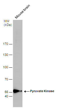
Pyruvate Kinase antibody detects Pyruvate Kinase protein by western blot analysis. Mouse tissue extracts (50 µg) was separated by 7.5% SDS-PAGE, and the membrane was blotted with Pyruvate Kinase antibody diluted by 1:1000. The HRP-conjugated anti-rabbit IgG antibody was used to detect the primary antibody.Application
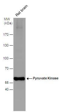
Pyruvate Kinase antibody detects Pyruvate Kinase protein by western blot analysis. Rat tissue extracts (50 µg) was separated by 7.5% SDS-PAGE, and the membrane was blotted with Pyruvate Kinase antibody diluted by 1:1000. The HRP-conjugated anti-rabbit IgG antibody was used to detect the primary antibody.Application
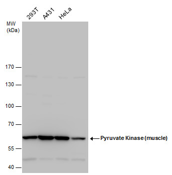
Pyruvate Kinase (muscle) antibody detects Pyruvate Kinase (muscle) protein by western blot analysis. Various whole cell extracts (30 µg) were separated by 7.5% SDS-PAGE, and blotted with Pyruvate Kinase (muscle) antibody diluted by 1:1000. The HRP-conjugated anti-rabbit IgG antibody was used to detect the primary antibody.Application
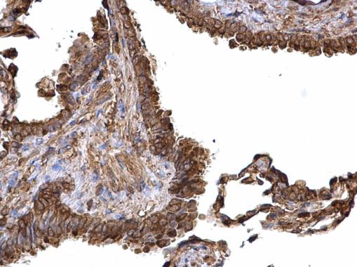
Pyruvate Kinase (muscle) antibody detects Pyruvate Kinase (muscle) protein at cytosol on human lung carcinoma by immunohistochemical analysis. Sample: Paraffin-embedded human lung carcinoma. Pyruvate Kinase (muscle) antibody dilution: 1:500.WB
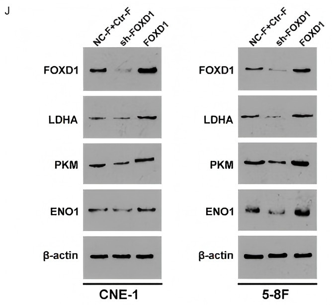
(J) FOXD1 knockdown induces the protein level of LDHA, PKM, and ENO1, while FOXD1 overexpression increases the expression level of these genes in CNE-1 and 5-8F cells.WB
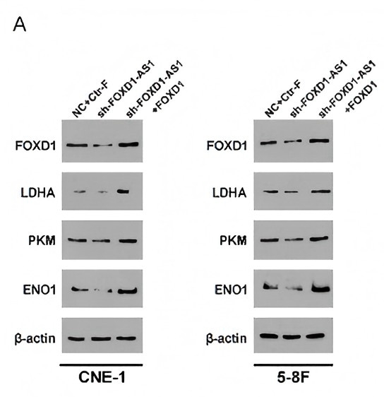
(A) Knockdown of FOXD1-AS1 could decrease the protein level of FOXD1, LDHA, PKM, and ENO1, and overexpression of FOXD1 significantly reverses the effects of FOXD1-AS1 silencing in CNE-1 and 5-8F cells.WB
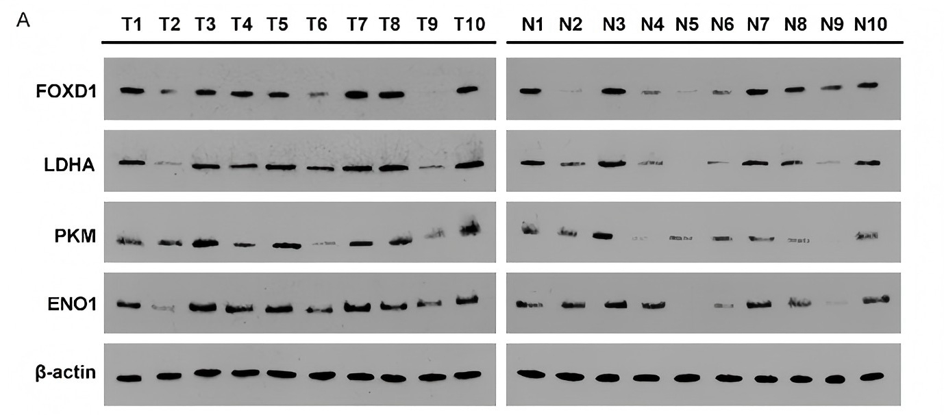
A. Western blotting analysis of FOXD1, LDHA, PKM, and ENO1 expression in 10 pairs of tumor tissues and normal tissues from patients with NPC.| Product Name | PKM antibody |
|---|---|
| Antibody Type | Primary Antibodies |
| Product description | Rabbit Polyclonal antibody to PKM (muscle) (pyruvate kinase, muscle)The protein encoded by this gene is a pyruvate kinase that catalyzes the production of phosphoenolpyruvate from pyruvate and ATP. This protein has been shown to interact with thyroid hormone, and thus may mediate cellular metabolic effects induced by thyroid hormones. This protein has been found to bind Opa protein, a bacterial outer membrane protein involved in gonococcal adherence to and invasion of human cells, suggesting a role of this protein in bacterial pathogenesis. Three alternatively spliced transcript variants encoding two distinct isoforms have been reported. [provided by RefSeq] |
| Immunogen | Recombinant protein encompassing a sequence within the center region of human Pyruvate Kinase (muscle). The exact sequence is proprietary. |
| Clonality | Polyclonal |
|---|---|
| Isotype | IgG |
| Host Species | Rabbit |
| Tested Applications | ICC/IFIHC-PIPWB |
| *Optimal dilutions/concentrations should be determined by the researcher. | |
| Species Reactivity | HumanMouseRat |
| Predicted Reactivity | Xenopus laevis, Rabbit, Chicken, BovineXenopus laevis (90%), Rabbit (97%), Chicken (83%), Bovine (97%) |
| Concentration | 1mg/ |
| Purification | Affinity purified |
| Gene Synonyms | CTHBP antibody, MGC3932 antibody, OIP3 antibody, PK3 antibody, PKM antibody, TCB antibody, THBP1 antibody, PKM2 antibody, PK, muscle type antibody, tumor M2-PK antibody, OIP-3 antibody, pyruvate kinase 2/3 antibody, pyruvate kinase muscle isozyme antibody, |
|---|---|
| Molecular Weight(MW) | 58 kDa (note) (9) |
| Cellular Localization | Cytoplasm , Nucleus |
Application
Non-transfected (–) and transfected (+) 293T whole cell extracts (30 µg) were separated by 7.5% SDS-PAGE, and the membrane was blotted with PKM antibody diluted at 1:5000. The HRP-conjugated anti-rabbit IgG antibody was used to detect the primary antibody.
Application
Immunoprecipitation of PKM2 protein from 293T whole cell extracts using 5 μg of Pyruvate Kinase (muscle) antibody . Western blot analysis was performed using Pyruvate Kinase (muscle) antibody . EasyBlot anti-Rabbit IgG was used as a secondary reagent.
Application
Pyruvate Kinase (muscle) antibody detects Pyruvate Kinase (muscle) protein by western blot analysis. Various whole cell extracts (30 µg) were separated by 10% SDS-PAGE, and the membrane was blotted with Pyruvate Kinase (muscle) antibody diluted by 1:1000. The HRP-conjugated anti-rabbit IgG antibody was used to detect the primary antibody.
Application
Pyruvate Kinase (muscle) antibody detects Pyruvate Kinase (muscle) protein at cytoplasm by immunofluorescent analysis. Sample: HeLa cells were fixed in 4% paraformaldehyde at RT for 15 min. Green: Pyruvate Kinase (muscle) protein stained by Pyruvate Kinase (muscle) antibody diluted at 1:500. Red: alpha Tubulin, a cytoskeleton marker, stained by alpha Tubulin antibody [GT114] diluted at 1:500. Blue: Hoechst 33342 staining.
Application
Pyruvate Kinase antibody detects Pyruvate Kinase protein by western blot analysis. Mouse tissue extracts (50 µg) was separated by 7.5% SDS-PAGE, and the membrane was blotted with Pyruvate Kinase antibody diluted by 1:1000. The HRP-conjugated anti-rabbit IgG antibody was used to detect the primary antibody.
Application
Pyruvate Kinase antibody detects Pyruvate Kinase protein by western blot analysis. Rat tissue extracts (50 µg) was separated by 7.5% SDS-PAGE, and the membrane was blotted with Pyruvate Kinase antibody diluted by 1:1000. The HRP-conjugated anti-rabbit IgG antibody was used to detect the primary antibody.
Application
Pyruvate Kinase (muscle) antibody detects Pyruvate Kinase (muscle) protein by western blot analysis. Various whole cell extracts (30 µg) were separated by 7.5% SDS-PAGE, and blotted with Pyruvate Kinase (muscle) antibody diluted by 1:1000. The HRP-conjugated anti-rabbit IgG antibody was used to detect the primary antibody.
Application
Pyruvate Kinase (muscle) antibody detects Pyruvate Kinase (muscle) protein at cytosol on human lung carcinoma by immunohistochemical analysis. Sample: Paraffin-embedded human lung carcinoma. Pyruvate Kinase (muscle) antibody dilution: 1:500.
WB
(J) FOXD1 knockdown induces the protein level of LDHA, PKM, and ENO1, while FOXD1 overexpression increases the expression level of these genes in CNE-1 and 5-8F cells.
WB
(A) Knockdown of FOXD1-AS1 could decrease the protein level of FOXD1, LDHA, PKM, and ENO1, and overexpression of FOXD1 significantly reverses the effects of FOXD1-AS1 silencing in CNE-1 and 5-8F cells.
WB
A. Western blotting analysis of FOXD1, LDHA, PKM, and ENO1 expression in 10 pairs of tumor tissues and normal tissues from patients with NPC.| Positive Control | 293T , A431 , HeLa , HepG2 , mouse brain , rat brain |
|---|---|
| Application Notes | *Optimal dilutions/concentrations should be determined by the researcher. |
| Form | Liquid |
|---|---|
| Storage Instructions | Keep as concentrated solution. Aliquot and store at -20ºC or below. Avoid multiple freeze-thaw cycles. |
| Storage Buffer | 1XPBS, 20% Glycerol (pH7). 0.025% ProClin 300 was added as a preservative. |


