Application
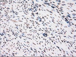
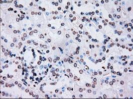
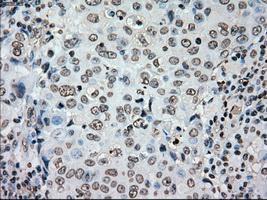
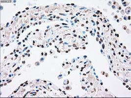
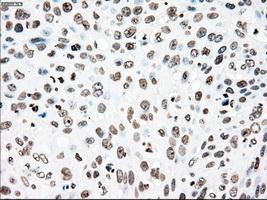
Immunohistochemical staining of paraffin-embedded endometrium tissue using anti-PLK1mouse monoclonal antibody. ( Dilution 1:50)Application
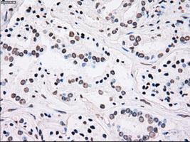
Immunohistochemical staining of paraffin-embedded Kidney tissue using anti-PLK1mouse monoclonal antibody. ( Dilution 1:50)Application
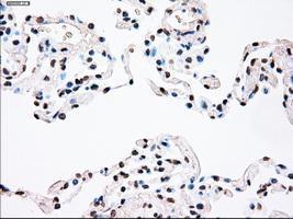
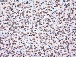
Immunohistochemical staining of paraffin-embedded lung tissue using anti-PLK1mouse monoclonal antibody. ( Dilution 1:50)Application
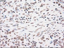
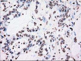
Immunohistochemical staining of paraffin-embedded Adenocarcinoma of breast tissue using anti-PLK1 mouse monoclonal antibody. ( Dilution 1:50)Application
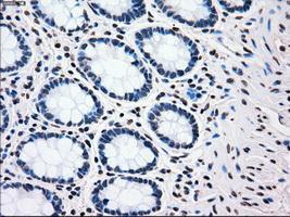
Immunohistochemical staining of paraffin-embedded colon tissue using anti-PLK1mouse monoclonal antibody. ( Dilution 1:50)Application
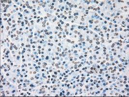
Immunohistochemical staining of paraffin-embedded pancreas tissue using anti-PLK1mouse monoclonal antibody. ( Dilution 1:50)Application
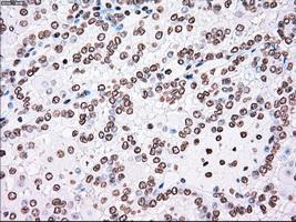
Immunohistochemical staining of paraffin-embedded Carcinoma of bladder tissue using anti-PLK1mouse monoclonal antibody. ( Dilution 1:50)Application
Immunohistochemical staining of paraffin-embedded Carcinoma of kidney tissue using anti-PLK1mouse monoclonal antibody. ( Dilution 1:50)Application
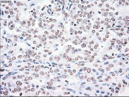
Immunohistochemical staining of paraffin-embedded Carcinoma of lung tissue using anti-PLK1mouse monoclonal antibody. ( Dilution 1:50)Application
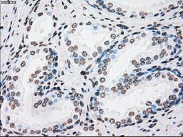
Immunohistochemical staining of paraffin-embedded prostate tissue using anti-PLK1mouse monoclonal antibody. ( Dilution 1:50)Application
Immunohistochemical staining of paraffin-embedded Adenocarcinoma of colon tissue using anti-PLK1mouse monoclonal antibody. ( Dilution 1:50)Application
Immunohistochemical staining of paraffin-embedded Adenocarcinoma of endometrium tissue using anti-PLK1mouse monoclonal antibody. ( Dilution 1:50)Application
Immunohistochemical staining of paraffin-embedded Carcinoma of pancreas tissue using anti-PLK1mouse monoclonal antibody. ( Dilution 1:50)Application
Immunohistochemical staining of paraffin-embedded Carcinoma of prostate tissue using anti-PLK1mouse monoclonal antibody. ( Dilution 1:50)Application
Immunohistochemical staining of paraffin-embedded Carcinoma of thyroid tissue using anti-PLK1mouse monoclonal antibody. ( Dilution 1:50)Application
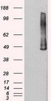
HEK293T cells were transfected with the pCMV6-ENTRY control (Left lane) or pCMV6-ENTRY PLK1 (Right lane) cDNA for 48 hrs and lysed. Equivalent amounts of cell lysates (5 ug per lane) were separated by SDS-PAGE and immunoblotted with anti-PLK1.Application
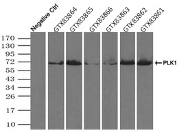
Immunoprecipitation (IP) of PLK1 by using TrueMab monoclonal anti-PLK1 antibodies (Negative control: IP without adding anti-PLK1 antibody.). For each experiment, 500ul of DDK tagged PLK1 overexpression lysates (at 1:5 dilution with HEK293T lysate), 2ug of anti-PLK1 antibody and 20ul (0.1mg) of goat anti-mouse conjugated magnetic beads were mixed and incubated overnight. After extensive wash to remove any non-specific binding, the immuno-precipitated products were analyzed with rabbit anti-DDK polyclonal antibody.| Product Name | PLK1 antibody [1D4] |
|---|---|
| Antibody Type | Primary Antibodies |
| Product description | Mouse monoclonal antibody [1D4] to PLK1Serine/threonine-protein kinase that performs several important functions throughout M phase of the cell cycle, including the regulation of centrosome maturation and spindle assembly, the removal of cohesins from chromosome arms, the inactivation of APC/C inhibitors, and the regulation of mitotic exit and cytokinesis. Required for recovery after DNA damage checkpoint and entry into mitosis. Required for kinetochore localization of BUB1B. Phosphorylates SGOL1. Required for spindle pole localization of isoform 3 of SGOL1 and plays a role in regulating its centriole cohesion function. Phosphorylates BORA, and thereby promotes the degradation of BORA. Contributes to the regulation of AURKA function. Regulates TP53 stability through phosphorylation of TOPORS. |
| Immunogen | Protein expressed in 293T cell transfected with human PLK1 expression vector |
| Clonality | Monoclonal |
|---|---|
| Isotype | IgG2 |
| Clone Number | 1D4 |
| Host Species | Mouse |
| Tested Applications | IHC,IHC-P,IP,WB |
| Optimal working dilutions should be determined experimentally by the end user. | |
| Species Reactivity | Human |
| Concentration | 1mg/ml |
| Purification | Affinity purified |
| Gene Synonyms | PLK, PLK1, STPK13 |
|---|---|
| Target | PLK1 |
| Tissue Specificity | is specific for human PLK1. |
| Cellular Localization | During early stages of mitosis, the phosphorylated form is detected on centrosomes and kinetochores. Localizes to the outer kinetochore. Presence of SGOL1 and interaction with the phosphorylated form of BUB1 is required for the kinetochore localization. |
Application
Immunohistochemical staining of paraffin-embedded endometrium tissue using anti-PLK1mouse monoclonal antibody. ( Dilution 1:50)
Application
Immunohistochemical staining of paraffin-embedded Kidney tissue using anti-PLK1mouse monoclonal antibody. ( Dilution 1:50)
Application
Immunohistochemical staining of paraffin-embedded lung tissue using anti-PLK1mouse monoclonal antibody. ( Dilution 1:50)
Application
Immunohistochemical staining of paraffin-embedded Adenocarcinoma of breast tissue using anti-PLK1 mouse monoclonal antibody. ( Dilution 1:50)
Application
Immunohistochemical staining of paraffin-embedded colon tissue using anti-PLK1mouse monoclonal antibody. ( Dilution 1:50)
Application
Immunohistochemical staining of paraffin-embedded pancreas tissue using anti-PLK1mouse monoclonal antibody. ( Dilution 1:50)
Application
Immunohistochemical staining of paraffin-embedded Carcinoma of bladder tissue using anti-PLK1mouse monoclonal antibody. ( Dilution 1:50)
Application
Immunohistochemical staining of paraffin-embedded Carcinoma of kidney tissue using anti-PLK1mouse monoclonal antibody. ( Dilution 1:50)
Application
Immunohistochemical staining of paraffin-embedded Carcinoma of lung tissue using anti-PLK1mouse monoclonal antibody. ( Dilution 1:50)
Application
Immunohistochemical staining of paraffin-embedded prostate tissue using anti-PLK1mouse monoclonal antibody. ( Dilution 1:50)
Application
Immunohistochemical staining of paraffin-embedded Adenocarcinoma of colon tissue using anti-PLK1mouse monoclonal antibody. ( Dilution 1:50)
Application
Immunohistochemical staining of paraffin-embedded Adenocarcinoma of endometrium tissue using anti-PLK1mouse monoclonal antibody. ( Dilution 1:50)
Application
Immunohistochemical staining of paraffin-embedded Carcinoma of pancreas tissue using anti-PLK1mouse monoclonal antibody. ( Dilution 1:50)
Application
Immunohistochemical staining of paraffin-embedded Carcinoma of prostate tissue using anti-PLK1mouse monoclonal antibody. ( Dilution 1:50)
Application
Immunohistochemical staining of paraffin-embedded Carcinoma of thyroid tissue using anti-PLK1mouse monoclonal antibody. ( Dilution 1:50)
Application
HEK293T cells were transfected with the pCMV6-ENTRY control (Left lane) or pCMV6-ENTRY PLK1 (Right lane) cDNA for 48 hrs and lysed. Equivalent amounts of cell lysates (5 ug per lane) were separated by SDS-PAGE and immunoblotted with anti-PLK1.
Application
Immunoprecipitation (IP) of PLK1 by using TrueMab monoclonal anti-PLK1 antibodies (Negative control: IP without adding anti-PLK1 antibody.). For each experiment, 500ul of DDK tagged PLK1 overexpression lysates (at 1:5 dilution with HEK293T lysate), 2ug of anti-PLK1 antibody and 20ul (0.1mg) of goat anti-mouse conjugated magnetic beads were mixed and incubated overnight. After extensive wash to remove any non-specific binding, the immuno-precipitated products were analyzed with rabbit anti-DDK polyclonal antibody.| Application Notes | Optimal working dilutions should be determined experimentally by the end user. |
|---|
| Form | Liquid |
|---|---|
| Storage Instructions | For short-term storage, store at 4°C or aliquot into working amounts and store at -20°C. For long-term storage, store at -70°C (aliquotted). Avoid repeated freeze-thaw cycles. |
| Storage Buffer | PBS (PH 7.3) containing 1% BSA, 50% glycerol and 0.02% sodium azide. |


