Application
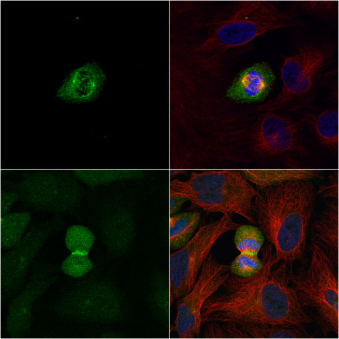
PLK1 antibody [N2C2], Internal detects PLK1 protein at midbody by immunofluorescent analysis. Sample: HeLa cells were fixed in 2% paraformadehyde at RT for 30 min. Green: PLK1 protein stained by PLK1 antibody [N2C2], Internal diluted at 1:1000. Red: alpha Tubulin, a cytoskeleton marker, stained by alpha Tubulin antibody [GT114] diluted at 1:1000. Blue: Hoechst 33342 staining.Application
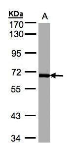
Sample(30 µg whole cell lysate) A:Raji 7.5% SDS PAGE diluted at 1:1000 The HRP-conjugated anti-rabbit IgG antibody was used to detect the primary antibody.Application
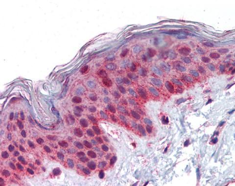
Immunohistochemical analysis of paraffin-embedded human skin, using PLK1 antibody(10 μg/ml).| Product Name | PLK1 antibody , Internal |
|---|---|
| Antibody Type | Primary Antibodies |
| Product description | Rabbit Polyclonal antibody to PLK1 (polo-like kinase 1)Serine/threonine-protein kinase that performs several important functions throughout M phase of the cell cycle, including the regulation of centrosome maturation and spindle assembly, the removal of cohesins from chromosome arms, the inactivation of APC/C inhibitors, and the regulation of mitotic exit and cytokinesis. Required for recovery after DNA damage checkpoint and entry into mitosis. Required for kinetochore localization of BUB1B. Phosphorylates SGOL1. Required for spindle pole localization of isoform 3 of SGOL1 and plays a role in regulating its centriole cohesion function. Phosphorylates BORA, and thereby promotes the degradation of BORA. Contributes to the regulation of AURKA function. Regulates TP53 stability through phosphorylation of TOPORS. |
| Immunogen | Recombinant protein encompassing a sequence within the center region of human PLK1. The exact sequence is proprietary. |
| Clonality | Polyclonal |
|---|---|
| Isotype | IgG |
| Host Species | Rabbit |
| Tested Applications | ICC/IF,IHC-P,WB |
| *Optimal dilutions/concentrations should be determined by the researcher. | |
| Species Reactivity | Human |
| Predicted Reactivity | Mouse, Rat, Xenopus laevis, Chicken, BovineMouse (95%), Rat (95%), Xenopus laevis (80%), Chicken (80%), Bovine (95%) |
| Concentration | 1mg/ml (Please refer to the vial label for the specific concentration) |
| Purification | Affinity purified |
| Gene Synonyms | PLK antibody, STPK13 antibody, PLK1 antibody, serine/threonine-protein kinase 13 antibody, polo (Drosophia)-like kinase antibody, polo like kinase antibody, cell cycle regulated protein kinase antibody, serine/threonine-protein kinase PLK1 antibody, PLK-1 |
|---|---|
| Molecular Weight(MW) | 68 kDa (note) |
| Cellular Localization | Nucleus. This antibody also stains PLK1 on spindles, kinetochores, and midbodies. |
Application
PLK1 antibody [N2C2], Internal detects PLK1 protein at midbody by immunofluorescent analysis. Sample: HeLa cells were fixed in 2% paraformadehyde at RT for 30 min. Green: PLK1 protein stained by PLK1 antibody [N2C2], Internal diluted at 1:1000. Red: alpha Tubulin, a cytoskeleton marker, stained by alpha Tubulin antibody [GT114] diluted at 1:1000. Blue: Hoechst 33342 staining.
Application
Sample(30 µg whole cell lysate) A:Raji 7.5% SDS PAGE diluted at 1:1000 The HRP-conjugated anti-rabbit IgG antibody was used to detect the primary antibody.
Application
Immunohistochemical analysis of paraffin-embedded human skin, using PLK1 antibody(10 μg/ml).| Application Notes | *Optimal dilutions/concentrations should be determined by the researcher. |
|---|
| Form | Liquid |
|---|---|
| Storage Instructions | Keep as concentrated solution. Aliquot and store at -20ºC or below. Avoid multiple freeze-thaw cycles. |
| Storage Buffer | 0.1M Tris, 0.1M Glycine, 10% Glycerol (pH7). 0.01% Thimerosal was added as a preservative. |


