Application
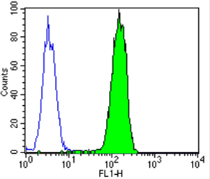
Flow cytometry analysis of PMP70 showing staining in the cytoplasm of 293T cells (green) compared to an isotype control (blue). Cells were harvested, adjusted to a concentration of 1-5x10^6 cells/ml, fixed with 2% paraformaldehyde and washed with PBS. Cells were blocked with a 2% solution of BSA-PBS for 30 min at room temperature and incubated with a PMP70 polyclonal antibody at a dilution of 0.5 ug/test for 60 min at room temperature. Cells were then incubated for 40 min at room temperature in the dark using a Dylight 488-conjugated goat anti-mouse IgG (H+L) secondary antibody and re-suspended in PBS for FACS analysis.Application
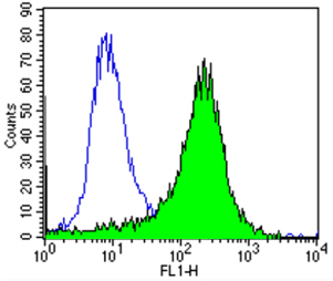
Flow cytometry analysis of PMP70 showing staining in the cytoplasm of HepG2 cells (green) compared to an isotype control (blue). Cells were harvested, adjusted to a concentration of 1-5x10^6 cells/ml, fixed with 2% paraformaldehyde and washed with PBS. Cells were blocked with a 2% solution of BSA-PBS for 30 min at room temperature and incubated with a PMP70 polyclonal antibody at a dilution of 0.5 ug/test for 60 min at room temperature. Cells were then incubated for 40 min at room temperature in the dark using a Dylight 488-conjugated goat anti-mouse IgG (H+L) secondary antibody and re-suspended in PBS for FACS analysis.| Product Name | PMP70 antibody |
|---|---|
| Antibody Type | Primary Antibodies |
| Product description | Rabbit Polyclonal antibody to PMP70Peroxisomes are single membrane organelles which are involved with many important biochemical pathways and are found in almost all eukaryotic cells. Various toxins including, phenols, alcohols, and aldehydes are targeted for modification by peroxisomes. Long chain fatty acid metabolism via beta-oxidation is also mediated by this organelle. Loss of peroxisomal functions may result in diseases such as Zellweger syndrome, hyperpipecolic acidemia, neonatal adrenoleukodystrophy, and infantile Refsum’s disease. Peroxisomal membrane protein 70 (PMP70) is an abundant, integral membrane protein of the peroxisome. This protein is induced by treatment with hypolipidemic agents in parallel with peroxisome proliferation and stimulation of the peroxisomal beta-oxidation enzymes. |
| Immunogen | Synthetic Peptide: C N(644) Y E F K K I T E D T V E F G S(659) |
| Clonality | Polyclonal |
|---|---|
| Host Species | Rabbit |
| Tested Applications | FACSICC/IFIHCWB |
| ICC/IF: 1:1,000 or 1:2,000: |
|
| Species Reactivity | HumanMouseRatHamsterPrimate |
| Concentration | 1mg/ml (Please refer to the vial label for the specific concentration) |
| Purification | Affinity purified |
| Gene Synonyms | Abcd3 AI313901 AU018866 AW146054 PMP68 PMP70 Pxmp1 ATP-binding cassette sub-family D (ALD) member 3 |
|---|---|
| Target | PMP70 |
Application
Flow cytometry analysis of PMP70 showing staining in the cytoplasm of 293T cells (green) compared to an isotype control (blue). Cells were harvested, adjusted to a concentration of 1-5x10^6 cells/ml, fixed with 2% paraformaldehyde and washed with PBS. Cells were blocked with a 2% solution of BSA-PBS for 30 min at room temperature and incubated with a PMP70 polyclonal antibody at a dilution of 0.5 ug/test for 60 min at room temperature. Cells were then incubated for 40 min at room temperature in the dark using a Dylight 488-conjugated goat anti-mouse IgG (H+L) secondary antibody and re-suspended in PBS for FACS analysis.
Application
Flow cytometry analysis of PMP70 showing staining in the cytoplasm of HepG2 cells (green) compared to an isotype control (blue). Cells were harvested, adjusted to a concentration of 1-5x10^6 cells/ml, fixed with 2% paraformaldehyde and washed with PBS. Cells were blocked with a 2% solution of BSA-PBS for 30 min at room temperature and incubated with a PMP70 polyclonal antibody at a dilution of 0.5 ug/test for 60 min at room temperature. Cells were then incubated for 40 min at room temperature in the dark using a Dylight 488-conjugated goat anti-mouse IgG (H+L) secondary antibody and re-suspended in PBS for FACS analysis.| Application Notes | ICC/IF: 1:1,000 or 1:2,000: |
|---|
| Form | Liquid |
|---|---|
| Storage Instructions | Keep as concentrated solution. Aliquot and store at -20ºC or below. Avoid multiple freeze-thaw cycles. |
| Storage Buffer | PBS containing 1 mg/ml BSA and 0.05% sodium azide. |


