Application
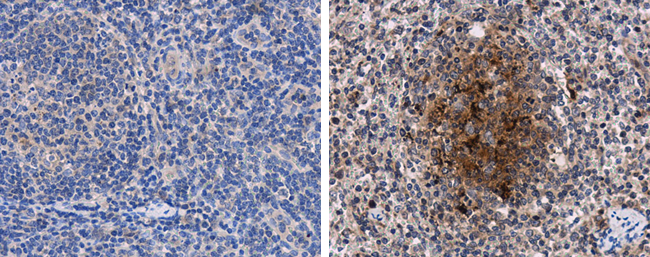
Porcine circovirus type 2 Capsid antibody detects PCV2 on lymph node by immunohistochemical analysis. Sample: Paraffin-embedded PCV2 free (left) and infected (right) swine lymph node. Porcine circovirus type 2 Capsid antibody dilution: 1:1000.Application
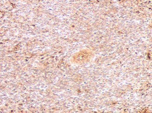
Porcine circovirus type 2 Capsid antibody didn't detects PCV2 at macrophages on spleen by immunohistochemical analysis. Sample: Paraffin-embedded PCV2 free swine spleen. Porcine circovirus type 2 Capsid antibody dilution: 1:500.Application
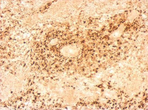
Porcine circovirus type 2 Capsid antibody detects PCV2 at macrophages on spleen by immunohistochemical analysis. Sample: Paraffin-embedded PCV2 infected swine spleen. Porcine circovirus type 2 Capsid antibody dilution: 1:500.Application
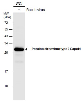
Lysate (30 µg) from Sf21 cells, expressing recombinant PCV2 protein by baculovirus system, was separated by 12% SDS-PAGE, and the membrane was blotted with Porcine circovirus type 2 Capsid antibody diluted at 1:500. The HRP-conjugated anti-rabbit IgG antibody was used to detect the primary antibody. (The lysate was provided courtesy of Dr. Wu lab at Chung Yuan Christian University, Department of Bioscience Technology.)Application
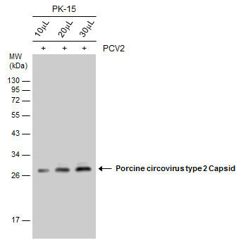
Infected (+) PK-15 whole cell extracts were separated by 12% SDS-PAGE, and the membrane was blotted with Porcine circovirus type 2 Capsid antibody diluted at 1:1000. The HRP-conjugated anti-rabbit IgG antibody was used to detect the primary antibody.| Product Name | Porcine circovirus type 2 Capsid antibody |
|---|---|
| Antibody Type | Primary Antibodies |
| Product description | Rabbit Polyclonal antibody to Porcine circovirus type 2 Capsid ProteinThe PCV2 virion measures approximately 17 nm in diameter, is non-enveloped and consists of a circular single-stranded DNA surrounded by an icosahedral capsid. The ambisense DNA molecule contains about 1.77 kb and 11 putative open reading frames (ORFs). PCV2 has been associated with a number of disease syndromes in pigs. Post-weaning multisystemic wasting syndrome (PMWS) is an important emerging viral disease affecting nursery and grower pigs, which has recently been associated with porcine circovirus type 2 (PCV2) infections. PMWS has been reported in Europe, South East Asia, and the Americas. |
| Clonality | Polyclonal |
|---|---|
| Isotype | IgG |
| Host Species | Rabbit |
| Tested Applications | IHC-PWB |
| *Optimal dilutions/concentrations should be determined by the researcher.
: |
|
| Species Reactivity | Porcine circovirus type 2 |
| Concentration | 1mg/ml (Please refer to the vial label for the specific concentration) |
| Purification | Affinity purified |
| Gene Synonyms | ORF2 |
|---|---|
| Molecular Weight(MW) | 28 kDa (note) |
Application
Porcine circovirus type 2 Capsid antibody detects PCV2 on lymph node by immunohistochemical analysis. Sample: Paraffin-embedded PCV2 free (left) and infected (right) swine lymph node. Porcine circovirus type 2 Capsid antibody dilution: 1:1000.
Application
Porcine circovirus type 2 Capsid antibody didn't detects PCV2 at macrophages on spleen by immunohistochemical analysis. Sample: Paraffin-embedded PCV2 free swine spleen. Porcine circovirus type 2 Capsid antibody dilution: 1:500.
Application
Porcine circovirus type 2 Capsid antibody detects PCV2 at macrophages on spleen by immunohistochemical analysis. Sample: Paraffin-embedded PCV2 infected swine spleen. Porcine circovirus type 2 Capsid antibody dilution: 1:500.
Application
Lysate (30 µg) from Sf21 cells, expressing recombinant PCV2 protein by baculovirus system, was separated by 12% SDS-PAGE, and the membrane was blotted with Porcine circovirus type 2 Capsid antibody diluted at 1:500. The HRP-conjugated anti-rabbit IgG antibody was used to detect the primary antibody. (The lysate was provided courtesy of Dr. Wu lab at Chung Yuan Christian University, Department of Bioscience Technology.)
Application
Infected (+) PK-15 whole cell extracts were separated by 12% SDS-PAGE, and the membrane was blotted with Porcine circovirus type 2 Capsid antibody diluted at 1:1000. The HRP-conjugated anti-rabbit IgG antibody was used to detect the primary antibody.| Positive Control | PCV2-infected PK-15 , PCV2 transfected |
|---|---|
| Application Notes | *Optimal dilutions/concentrations should be determined by the researcher.
: |
| Form | Liquid |
|---|---|
| Storage Instructions | Keep as concentrated solution. Aliquot and store at -20ºC or below. Avoid multiple freeze-thaw cycles. |
| Storage Buffer | 1XPBS, 20% Glycerol (pH7). 0.01% Thimerosal was added as a preservative. |


