Application
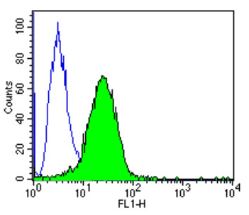
Flow cytometry analysis of PPAR alpha showing positive staining in the nucleus of 3T3 cells compared to an isotype control (blue). Cells were harvested, adjusted to a concentration of 1-5x10^6 cells/ml, fixed with 2% paraformaldehyde, washed with PBS, and incubated with PPAR alpha monoclonal antibody at a dilution of 0.25 ug/test for 60 min at room temperature. Cells were then blocked in a solution of 2% BSA-PBS for 30 min at room temperature, incubated for 40 min at room temperature in the dark using a Dylight 488-conjugated goat anti-mouse IgG (H+L) secondary antibody, and re-suspended in PBS for FACS analysis.Application
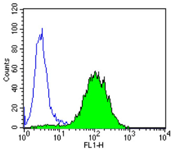
Flow cytometry analysis of PPAR alpha showing positive staining in the nucleus of Hela cells compared to an isotype control (blue). Cells were harvested, adjusted to a concentration of 1-5x10^6 cells/ml, fixed with 2% paraformaldehyde, washed with PBS, and incubated with PPAR alpha monoclonal antibody at a dilution of 0.25 ug/test for 60 min at room temperature. Cells were then blocked in a solution of 2% BSA-PBS for 30 min at room temperature, incubated for 40 min at room temperature in the dark using a Dylight 488-conjugated goat anti-mouse IgG (H+L) secondary antibody, and re-suspended in PBS for FACS analysis.Application
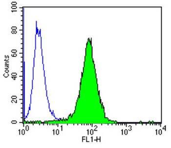
Flow cytometry analysis of PPAR alpha showing positive staining in the nucleus of Jurkat cells compared to an isotype control (blue). Cells were harvested, adjusted to a concentration of 1-5x10^6 cells/ml, fixed with 2% paraformaldehyde, washed with PBS, and incubated with PPAR alpha monoclonal antibody at a dilution of 0.25 ug/test for 60 min at room temperature. Cells were then blocked in a solution of 2% BSA-PBS for 30 min at room temperature, incubated for 40 min at room temperature in the dark using a Dylight 488-conjugated goat anti-mouse IgG (H+L) secondary antibody, and re-suspended in PBS for FACS analysis.Application
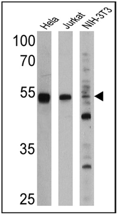
Western blot analysis of PPAR alpha was performed by loading 25 ug of Hela (Lane 1), Jurkat (Lane 2), and NIH-3T3 cell lysates (Lane 3) and a molecular weight protein ladder onto an SDS polyacrylamide gel. Proteins were transferred to a PVDF membrane and blocked with a blocking buffer at 4ºC overnight. The membrane was probed with a PPAR alpha monoclonal antibody at a dilution of 1:1000 (Hela and Jurkat) and 1:500 (NIH-3T3) overnight at 4°C, washed in TBST, and probed with an HRP-conjugated secondary antibody for 1 hr at room temperature in the dark. Chemiluminescent detection was performed. Results show a band at 52 kDa in all three cell lines.| Product Name | PPAR alpha antibody |
|---|---|
| Antibody Type | Primary Antibodies |
| Product description | Mouse Monoclonal antibody to PPAR alphaPeroxisome proliferators are non-genotoxic carcinogens which are purported to exert their effect on cells through their interaction with members of the nuclear hormone receptor family, termed peroxisome proliferator activated receptors (PPARs). Nuclear hormone receptors are ligand-dependent intracellular proteins that stimulate transcription of specific genes by binding to specific DNA sequences following activation by the appropriate ligand. Studies indicate that PPARs are activated by peroxisome proliferators such as clofibric acid, nafenopin, and WY-14,643, as well as by some fatty acids. It has also been shown that PPARs can induce transcription of acyl coenzyme A oxidase & cytochrome P450 A6 (CYP450 A6) through interaction with specific response elements. PPAR alpha is activated by free fatty acids including linoleic, arachidonic, and oleic acids. Induction of peroxisomes by this mechanism leads to a reduction in blood triglyceride levels. PPAR alpha is expressed mainly in skeletal muscle, heart, liver, and kidney and is thought to regulate many genes involved in the beta-oxidation of fatty acids. Activation of rat liver PPAR alpha has been shown to suppress hepatocyte apoptosis. PPAR alpha, like several other nuclear hormone receptors, heterodimerizes with retinoic X receptor (RXR) alpha to form a transcriptionally competent complex. The corresponding gene for the PPAR alpha is NR1C1. |
| Immunogen | Purified recombinant PPAR alpha protein. |
| Clonality | Monoclonal |
|---|---|
| Isotype | IgG2 |
| Clone Number | 3B6/PPAR |
| Host Species | Mouse |
| Tested Applications | FACSGSAIHCIPWBChIP |
| For WB: use at a concentration of 2-5 µg/ml. For IP: Assay dependent. Optimal dilutions / concentrations should be determined by the end user. By Western blot, this antibody detects a ~52 kDa protein which corresponds to PPAR alpha from mouse adFor IPose tissue extract.: |
|
| Species Reactivity | HumanMouseRatRabbitBovine |
| Concentration | 1mg/ml (Please refer to the vial label for the specific concentration) |
| Purification | Affinity purified |
| Gene Synonyms | PPARA MGC2237 MGC2452 NR1C1 PPAR PPARalpha hPPAR peroxisome proliferator-activated receptor alpha |
|---|---|
| Target | PPAR alpha |
Application
Flow cytometry analysis of PPAR alpha showing positive staining in the nucleus of 3T3 cells compared to an isotype control (blue). Cells were harvested, adjusted to a concentration of 1-5x10^6 cells/ml, fixed with 2% paraformaldehyde, washed with PBS, and incubated with PPAR alpha monoclonal antibody at a dilution of 0.25 ug/test for 60 min at room temperature. Cells were then blocked in a solution of 2% BSA-PBS for 30 min at room temperature, incubated for 40 min at room temperature in the dark using a Dylight 488-conjugated goat anti-mouse IgG (H+L) secondary antibody, and re-suspended in PBS for FACS analysis.
Application
Flow cytometry analysis of PPAR alpha showing positive staining in the nucleus of Hela cells compared to an isotype control (blue). Cells were harvested, adjusted to a concentration of 1-5x10^6 cells/ml, fixed with 2% paraformaldehyde, washed with PBS, and incubated with PPAR alpha monoclonal antibody at a dilution of 0.25 ug/test for 60 min at room temperature. Cells were then blocked in a solution of 2% BSA-PBS for 30 min at room temperature, incubated for 40 min at room temperature in the dark using a Dylight 488-conjugated goat anti-mouse IgG (H+L) secondary antibody, and re-suspended in PBS for FACS analysis.
Application
Flow cytometry analysis of PPAR alpha showing positive staining in the nucleus of Jurkat cells compared to an isotype control (blue). Cells were harvested, adjusted to a concentration of 1-5x10^6 cells/ml, fixed with 2% paraformaldehyde, washed with PBS, and incubated with PPAR alpha monoclonal antibody at a dilution of 0.25 ug/test for 60 min at room temperature. Cells were then blocked in a solution of 2% BSA-PBS for 30 min at room temperature, incubated for 40 min at room temperature in the dark using a Dylight 488-conjugated goat anti-mouse IgG (H+L) secondary antibody, and re-suspended in PBS for FACS analysis.
Application
Western blot analysis of PPAR alpha was performed by loading 25 ug of Hela (Lane 1), Jurkat (Lane 2), and NIH-3T3 cell lysates (Lane 3) and a molecular weight protein ladder onto an SDS polyacrylamide gel. Proteins were transferred to a PVDF membrane and blocked with a blocking buffer at 4ºC overnight. The membrane was probed with a PPAR alpha monoclonal antibody at a dilution of 1:1000 (Hela and Jurkat) and 1:500 (NIH-3T3) overnight at 4°C, washed in TBST, and probed with an HRP-conjugated secondary antibody for 1 hr at room temperature in the dark. Chemiluminescent detection was performed. Results show a band at 52 kDa in all three cell lines.| Application Notes | For WB: use at a concentration of 2-5 µg/ml. For IP: Assay dependent. Optimal dilutions / concentrations should be determined by the end user. By Western blot, this antibody detects a ~52 kDa protein which corresponds to PPAR alpha from mouse adFor IPose tissue extract.: |
|---|
| Form | Liquid |
|---|---|
| Storage Instructions | Keep as concentrated solution. Aliquot and store at -20ºC or below. Avoid multiple freeze-thaw cycles. |
| Storage Buffer | (1 mg/ml) in PBS containing 0.05% sodium azide. |


