Application
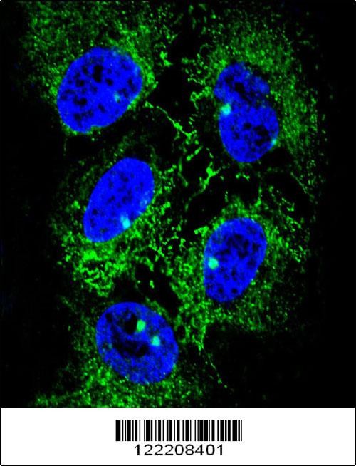
Confocal immunofluorescent analysis of ADH7 Antibody with NCI-H460 cell followed by Alexa Fluor 488-conjugated goat anti-rabbit lgG (green). DAPI was used to stain the cell nuclear (blue).Application
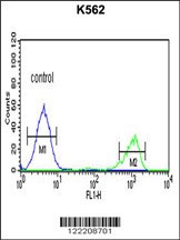
Flow cytometric analysis of K562 cells (right histogram) compared to a negative control cell (left histogram).FITC-conjugated goat-anti-rabbit secondary antibodies were used for the analysis.Application
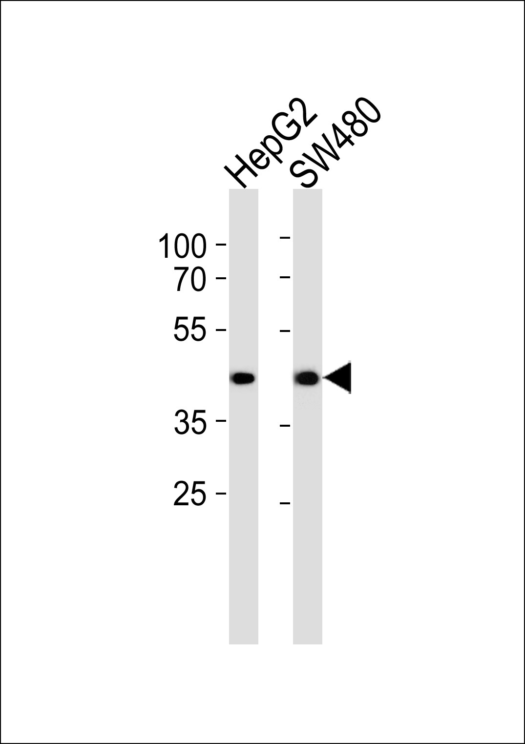
Western blot analysis of lysates from HepG2, SW480 cell line (from left to right), using ADH7 Antibody at 1:1000 at each lane.Application
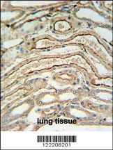
ADH7 Antibody IHC analysis in formalin fixed and paraffin embedded lung tissue followed by peroxidase conjugation of the secondary antibody and DAB staining.| Product Name | ADH7 Antibody |
|---|---|
| Antibody Type | Primary Antibodies |
| Antigen Alias | Alcohol dehydrogenase class 4 mu/sigma chain, Alcohol dehydrogenase class IV mu/sigma chain, Gastric alcohol dehydrogenase, Retinol dehydrogenase, ADH7 |
| Product description | This gene encodes class IV alcohol dehydrogenase 7 mu or sigma subunit, which is a member of the alcohol dehydrogenase family. Members of this family metabolize a wide variety of substrates, including ethanol, retinol, other aliphatic alcohols, hydroxysteroids, and lipid peroxidation products. The enzyme encoded by this gene is inefficient in ethanol oxidation, but is the most active as a retinol dehydrogenase; thus it may participate in the synthesis of retinoic acid, a hormone important for cellular differentiation. The expression of this gene is much more abundant in stomach than liver, thus differing from the other known gene family members.1) Kedishvili, N.Y., et al. J. Biol. Chem. 270(8):3625-3630(1995) |
| Immunogen | This ADH7 antibody is generated from rabbits immunized with a KLH conjugated synthetic peptide between 318-346 amino acids from the C-terminal region of human ADH7. |
| Clonality | Polyclonal |
|---|---|
| Isotype | Ig |
| Host Species | Rabbit |
| Tested Applications | FACSIFIHC-PWB |
| For WB starting dilution is: 1:1000 For IHC-P starting dilution is: 1:50~100 For FACS starting dilution is: 1:10~50 For IF starting dilution is: 1:10~50: |
|
| Species Reactivity | Human |
| Concentration | 1mg/ml |
| Purification | Affinity purified |
| Gene Symbol | ADH7 |
|---|---|
| Alternative Names | Alcohol dehydrogenase class 4 mu/sigma chain Alcohol dehydrogenase class IV mu/sigma chain Gastric alcohol dehydrogenase Retinol dehydrogenase ADH7 |
| Molecular Weight(MW) | 41 kDa |
Application
Confocal immunofluorescent analysis of ADH7 Antibody with NCI-H460 cell followed by Alexa Fluor 488-conjugated goat anti-rabbit lgG (green). DAPI was used to stain the cell nuclear (blue).
Application
Flow cytometric analysis of K562 cells (right histogram) compared to a negative control cell (left histogram).FITC-conjugated goat-anti-rabbit secondary antibodies were used for the analysis.
Application
Western blot analysis of lysates from HepG2, SW480 cell line (from left to right), using ADH7 Antibody at 1:1000 at each lane.
Application
ADH7 Antibody IHC analysis in formalin fixed and paraffin embedded lung tissue followed by peroxidase conjugation of the secondary antibody and DAB staining.| Application Notes | For WB starting dilution is: 1:1000 For IHC-P starting dilution is: 1:50~100 For FACS starting dilution is: 1:10~50 For IF starting dilution is: 1:10~50: |
|---|
| Form | Liquid |
|---|---|
| Storage Instructions | Store at 4˚C for three months and -20˚C, stable for up to one year. As with all antibodies care should be taken to avoid repeated freeze thaw cycles. Antibodies should not be exposed to prolonged high temperatures. |
| Storage Buffer | Supplied in PBS with 0.09% (W/V) sodium azide. |


