Application
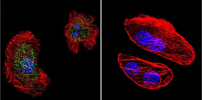
Immunofluorescent analysis of Phosphorylated Progesterone Receptor in U251 cells. Ser294-phosphorylated Progesterone Receptor staining (green), F-Actin staining with Phalloidin (red) and nuclei with DAPI (blue) is shown. Cells were grown on slides and fixed with formaldehyde prior to staining. Cells were probed without (control) or with Progesterone Receptor (phospho Ser294) antibody [608] at a dilution of 1:20 over night at 4 °C, washed with PBS and incubated with a proper secondary antibody. Images were taken at 60X magnification.| Product Name | Progesterone Receptor (phospho Ser294) antibody |
|---|---|
| Antibody Type | Primary Antibodies |
| Product description | Mouse monoclonal antibody [608] to Ser294-phosphorylated Progesterone ReceptorThis gene encodes a member of the steroid receptor superfamily. The encoded protein mediates the physiological effects of progesterone, which plays a central role in reproductive events associated with the establishment and maintenance of pregnancy. This gene uses two distinct promotors and translation start sites in the first exon to produce two isoforms, A and B. The two isoforms are identical except for the additional 165 amino acids found in the N-terminus of isoform B and mediate their own response genes and physiologic effects with little overlap. [provided by RefSeq, Jan 2011] |
| Immunogen | Synthetic phosphopeptide corresponding to residues P(288) M A P G R S(p) P L A T T V(300) of human Progesterone Receptor. |
| Modification | p-Ser294 |
| Clonality | Monoclonal |
|---|---|
| Isotype | IgG1 |
| Clone Number | 608 |
| Host Species | Mouse |
| Tested Applications | ELISAGSAICC/IFIHCIHC-PIPWB |
| IHC-P: 1:500: |
|
| Species Reactivity | HumanMouseRat |
| Concentration | 1mg/ml (Please refer to the vial label for the specific concentration) |
| Gene Synonyms | PGR progesterone receptor PR NR3C3 |
|---|---|
| Target | Phospho-Progesterone Rec. pSer294 |
Application
Immunofluorescent analysis of Phosphorylated Progesterone Receptor in U251 cells. Ser294-phosphorylated Progesterone Receptor staining (green), F-Actin staining with Phalloidin (red) and nuclei with DAPI (blue) is shown. Cells were grown on slides and fixed with formaldehyde prior to staining. Cells were probed without (control) or with Progesterone Receptor (phospho Ser294) antibody [608] at a dilution of 1:20 over night at 4 °C, washed with PBS and incubated with a proper secondary antibody. Images were taken at 60X magnification.| Application Notes | IHC-P: 1:500: |
|---|
| Form | Liquid |
|---|---|
| Storage Instructions | -20° C, Avoid Freeze/Thaw Cycles |
| Storage Buffer | Contains 0.05 % sodium azide and 1 mg/ml BSA |


