Application
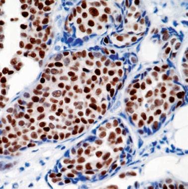
IHC Image using - Detection of Profesterone receptor by IHC in Human Breast Carcinoma| Product Name | Progesterone Receptor antibody |
|---|---|
| Antibody Type | Primary Antibodies |
| Product description | Mouse monoclonal [hpra 2 + hpra 3] to Progesterone ReceptorThe human Progesterone Receptor (PgR) is a member of the steroid family of nuclear receptors. PgR is found as a 120 kDa protein (Form B) or a 94 kDa protein (Form A) due to the use of alternative translation initiation sites. In its inactive state, PgR forms a multiprotein complex which includes heat shock proteins and immunophins. Upon binding of progesterone hormone to its receptor, there is a conformational change that allows dimerization and binding of the receptor to progesterone response elements (PRE) sequences, resulting in activated transcription. When bound to DNA, PgR can positively or negatively regulate gene transcription through the recruitment of coactivator (e.g. SRC 1) or corepressor (e.g. NCoR) proteins. PgR is phosphorylated on multiple serine residues through distinct groups of sites regulated by hormone and different kinases.Progesterone receptor is present in the CNS in monoenzymatic aromatic L-amino acid decarboxylase (AADC) containing cells of the preoptic area and also in neurons expressing both AADC and tyrosine hydroxylase (TH) in the arcuate nucleus. |
| Immunogen | BALB/C mice were injected with progesterone receptor from human endometrial carcinoma (EnCa 101) grown in athymic mice. |
| Clonality | Monoclonal |
|---|---|
| Isotype | IgG1 |
| Clone Number | hpra 2 + hpra 3 |
| Host Species | Mouse |
| Tested Applications | IHCIHC-P |
| IHC: 1/50 (30 min at RT). IHC-P: 1/50 (30 min at RT). Perform heat mediated antigen retrieval before commencing with IHC staining protocol. Optimal dilutions/concentrations should be determined by the end user.: |
|
| Species Reactivity | Human |
| Concentration | 1mg/ml |
| Gene Synonyms | PGR 607311 Progesterone receptor P06401 PR NR3C3 5241 |
|---|---|
| Target | Progesterone receptor |
| Tissue Specificity | reacts with two forms of human progesterone receptor, the 116 kD (triplet;hormone binding high) and 81kD (singlet; hormone binding low) molecular weight proteins. |
| Cellular Localization | Nuclear |
Application
IHC Image using - Detection of Profesterone receptor by IHC in Human Breast Carcinoma| Positive Control | Progesterone receptor (PR) expression is found in AADC containing cells in the CNS preoptic area and also arcuate nucleus neurons expressing AADC and TH. Uterus and Breast carcinoma tissue can also be used as positive controls. |
|---|---|
| Application Notes | IHC: 1/50 (30 min at RT). IHC-P: 1/50 (30 min at RT). Perform heat mediated antigen retrieval before commencing with IHC staining protocol. Optimal dilutions/concentrations should be determined by the end user.: |
| Form | Liquid |
|---|---|
| Storage Instructions | Keep as concentrated solution, aliquot and store at 4°C. Do not freeze. |
| Storage Buffer | Preservative: Sodium Azide |


