Application
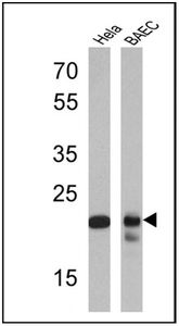
WB analysis of HeLa and BAEC lysates (25 ug) using Proteasome 20S Y antibody at a dilution of 1:1,000.Application
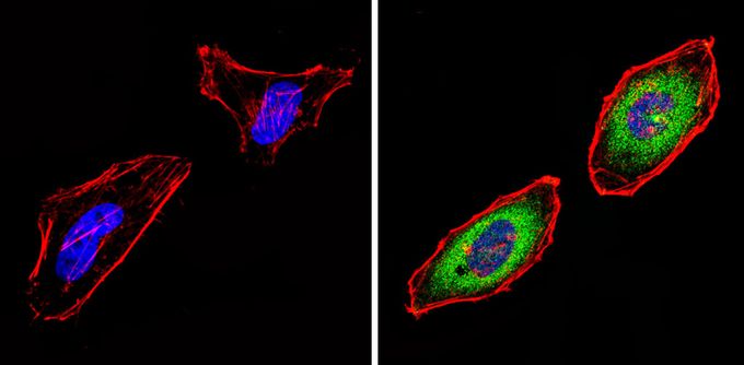
ICC/IF analysis of HeLa cells with (right) or without (left) Proteasome 20S Y antibody at a dilution of 1:20 (green). F-actin (red) was stained with a flourescent red phalloidin and nuclei (blue) were stained with DAPI. Images were taken at a magnification of 60x.Application
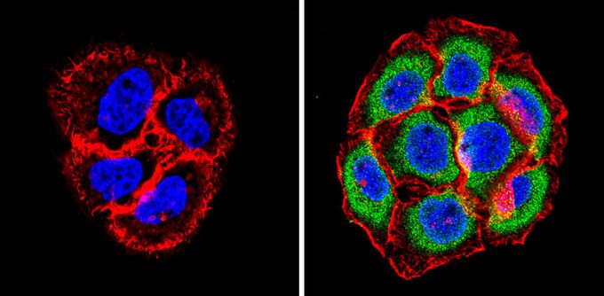
ICC/IF analysis of A431 cells with (right) or without (left) Proteasome 20S Y antibody at a dilution of 1:20 (green). F-actin (red) was stained with a flourescent red phalloidin and nuclei (blue) were stained with DAPI. Images were taken at a magnification of 60x.Application
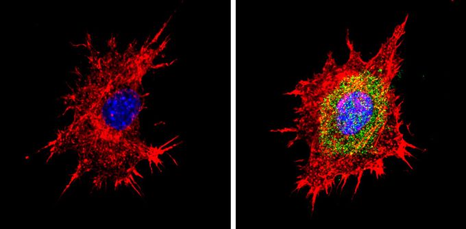
ICC/IF analysis of BAEC cells with (right) or without (left) Proteasome 20S Y antibody at a dilution of 1:20 (green). F-actin (red) was stained with a flourescent red phalloidin and nuclei (blue) were stained with DAPI. Images were taken at a magnification of 60x.| Product Name | Proteasome 20S Y antibody |
|---|---|
| Antibody Type | Primary Antibodies |
| Product description | Rabbit Polyclonal antibody to Proteasome 20S YProteolytic degradation is critical to the maintenance of appropriate levels of short-lived and regulatory proteins as important and diverse as those involved in cellular metabolism, heat shock and stress response, antigen presentation, modulation of cell surface receptors and ion channels, cell cycle regulation, transcription, and signalling factors. The ubiquitin-proteasome pathway deconstructs most proteins in the eukaryotic cell cytosol and nucleus. Other proteins are degraded via the vacuolar pathway which includes endosomes, lysosomes, and the endoplasmic reticulum. The 26S proteasome is an ATP-dependent, multisubunit (~31), barrel-shaped molecular machine with an apparent molecular weight of ~2.5 MDa. It consists of a 20S proteolytic core complex which is crowned at one or both ends by 19S regulatory subunit complexes. The 19S regulatory subunits recognize ubiquitinated proteins and play an essential role in unfolding and translocating targets into the lumen of the 20S subunit. The PA28/11S REG Activator protein complex functions as a proteolytic activator. This complex, consisting of alpha, beta, and gamma subunits, enhances the activity of the 20S proteolytic core. An enzymatic cascade is responsible for the attachment of multiple ubiquitin molecules to lysine residues of proteins targeted for degradation. Several genetic diseases are associated with defects in the ubiquitin-proteasome pathway. Some examples of affected proteins include those linked to cystic fibrosis (CF transmembrane regulator), Angelman’s syndrome (E6-AP), and Liddle syndrome (endothelial sodium channels). |
| Immunogen | Synthetic Peptides: C(41) R S G S A A D T Q A V/I A D A V T Y(58) |
| Clonality | Polyclonal |
|---|---|
| Host Species | Rabbit |
| Tested Applications | ICC/IFWB |
| ICC/IF: 1:10 - 1:100: |
|
| Species Reactivity | HumanMouseBovine |
| Concentration | 1mg/ml (Please refer to the vial label for the specific concentration) |
| Purification | Affinity purified |
| Gene Synonyms | Psmb6 Lmp19 Mpnd proteasome (prosome macropain) subunit beta type 6 |
|---|---|
| Target | Proteasome 20S Y |
Application
WB analysis of HeLa and BAEC lysates (25 ug) using Proteasome 20S Y antibody at a dilution of 1:1,000.
Application
ICC/IF analysis of HeLa cells with (right) or without (left) Proteasome 20S Y antibody at a dilution of 1:20 (green). F-actin (red) was stained with a flourescent red phalloidin and nuclei (blue) were stained with DAPI. Images were taken at a magnification of 60x.
Application
ICC/IF analysis of A431 cells with (right) or without (left) Proteasome 20S Y antibody at a dilution of 1:20 (green). F-actin (red) was stained with a flourescent red phalloidin and nuclei (blue) were stained with DAPI. Images were taken at a magnification of 60x.
Application
ICC/IF analysis of BAEC cells with (right) or without (left) Proteasome 20S Y antibody at a dilution of 1:20 (green). F-actin (red) was stained with a flourescent red phalloidin and nuclei (blue) were stained with DAPI. Images were taken at a magnification of 60x.| Application Notes | ICC/IF: 1:10 - 1:100: |
|---|
| Form | Liquid |
|---|---|
| Storage Instructions | Keep as concentrated solution. Aliquot and store at -20ºC or below. Avoid multiple freeze-thaw cycles. |
| Storage Buffer | PBS containing 1 mg/ml BSA and 0.05% sodium azide. |


