Application
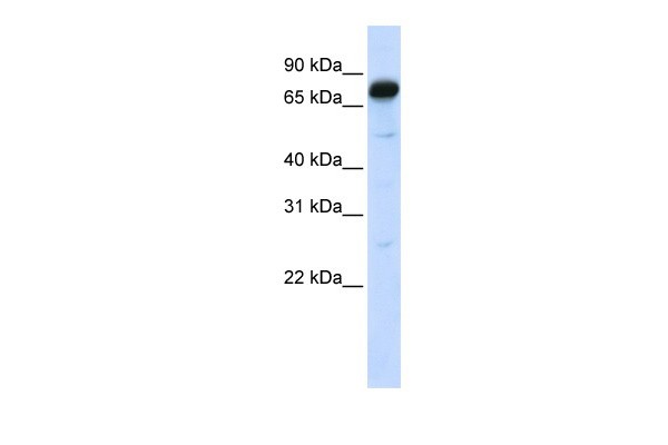
Antibody used in WB on Human 293T at 0.2-1 ug/ml.| Product Name | AMOT Antibody |
|---|---|
| Antibody Type | Primary Antibodies |
| Antigen Alias | AMOT, KIAA1071 |
| Product description | AMOT belongs to the motin family of angiostatin binding proteins characterized by conserved coiled-coil domains and C-terminal PDZ binding motifs. The protein is expressed predominantly in endothelial cells of capillaries as well as larger vessels of the placenta where it may mediate the inhibitory effect of angiostatin on tube formation and the migration of endothelial cells toward growth factors during the formation of new blood vessels. Alternative splicing results in multiple transcript variants encoding different isoforms.This gene belongs to the motin family of angiostatin binding proteins characterized by conserved coiled-coil domains and C-terminal PDZ binding motifs. The encoded protein is expressed predominantly in endothelial cells of capillaries as well as larger vessels of the placenta where it may mediate the inhibitory effect of angiostatin on tube formation and the migration of endothelial cells toward growth factors during the formation of new blood vessels. Alternative splicing results in multiple transcript variants encoding different isoforms.1) Holmgren, L., (2006) Proc. Natl. Acad. Sci. U.S.A. 103 (24), 9208-9213. |
| Immunogen | Antibody produced in rabbits immunized with a synthetic peptide corresponding a region of human AMOT. |
| Clonality | Polyclonal |
|---|---|
| Host Species | Rabbit |
| Tested Applications | ELISAWB |
| AMOT antibody can be used for detection of AMOT by ELISA at 1:1562500. AMOT antibody can be used for detection of AMOT by western blot at 1 μg/mL, and HRP conjugated secondary antibody should be diluted 1:50,000 - 100,000.: |
|
| Species Reactivity | Human |
| Concentration | 1mg/ml |
| Purification | Affinity purified |
| Gene Symbol | AMOT |
|---|---|
| Alternative Names | AMOT KIAA1071 |
| Molecular Weight(MW) | 72 kDa |
Application
Antibody used in WB on Human 293T at 0.2-1 ug/ml.| Positive Control | 1) 293T Cell Lysate |
|---|---|
| Application Notes | AMOT antibody can be used for detection of AMOT by ELISA at 1:1562500. AMOT antibody can be used for detection of AMOT by western blot at 1 μg/mL, and HRP conjugated secondary antibody should be diluted 1:50,000 - 100,000.: |
| Form | Liquid |
|---|---|
| Storage Instructions | For short periods of storage (days) store at 4˚C. For longer periods of storage, store AMOT antibody at -20˚C. As with any antibody avoid repeat freeze-thaw cycles. |
| Storage Buffer | Antibody is lyophilized in PBS buffer with 2% sucrose. Add 50 μL of distilled water. Final antibody concentration is 1 mg/mL. |


