Application
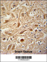
ARPC1A Antibody immunohistochemistry analysis in formalin fixed and paraffin embedded human brain tissue followed by peroxidase conjugation of the secondary antibody and DAB staining.Application
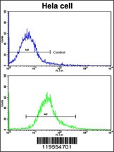
Flow cytometric analysis of hela cells using ARPC1A Antibody (bottom histogram) compared to a negative control cell (top histogram). FITC-conjugated goat-anti-rabbit secondary antibodies were used for the analysis.Application
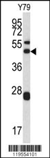
Western blot analysis of ARPC1A antibody in Y79 cell line lysates (35ug/lane)| Product Name | ARPC1A Antibody |
|---|---|
| Antibody Type | Primary Antibodies |
| Antigen Alias | Actin-related protein 2/3 complex subunit 1A, SOP2-like protein, ARPC1A, SOP2L |
| Product description | ARPC1A is one of seven subunits of the human Arp2/3 protein complex. This subunit is a member of the SOP2 family of proteins and is most similar to the protein ARPC1B. The similarity between these two proteins suggests that they both may function as p41 subunit of the human Arp2/3 complex that has been implicated in the control of actin polymerization in cells. It is possible that the p41 subunit is involved in assembling and maintaining the structure of the Arp2/3 complex. Multiple versions of the p41 subunit may adapt the functions of the complex to different cell types or developmental stages.1) Laurila,E., Genes Chromosomes Cancer 48 (4), 330-339 (2009) |
| Immunogen | This ARPC1A antibody is generated from rabbits immunized with a KLH conjugated synthetic peptide between 157-184 amino acids from the Central region of human ARPC1A. |
| Clonality | Polyclonal |
|---|---|
| Isotype | Ig |
| Host Species | Rabbit |
| Tested Applications | FACSIHC-PWB |
| For WB starting dilution is: 1:1000 For FACS starting dilution is: 1:10~50 For IHC-P starting dilution is: 1:50~100: |
|
| Species Reactivity | Human |
| Concentration | 1mg/ml |
| Gene Symbol | ARPC1A |
|---|---|
| Alternative Names | Actin-related protein 2/3 complex subunit 1A SOP2-like protein ARPC1A SOP2L |
| Molecular Weight(MW) | 42 kDa |
| Sequence Similarities | Predicted species reactivity based on immunogen sequence: Bovine |
Application
ARPC1A Antibody immunohistochemistry analysis in formalin fixed and paraffin embedded human brain tissue followed by peroxidase conjugation of the secondary antibody and DAB staining.
Application
Flow cytometric analysis of hela cells using ARPC1A Antibody (bottom histogram) compared to a negative control cell (top histogram). FITC-conjugated goat-anti-rabbit secondary antibodies were used for the analysis.
Application
Western blot analysis of ARPC1A antibody in Y79 cell line lysates (35ug/lane)| Application Notes | For WB starting dilution is: 1:1000 For FACS starting dilution is: 1:10~50 For IHC-P starting dilution is: 1:50~100: |
|---|
| Form | Liquid |
|---|---|
| Storage Instructions | Store at 4˚C for three months and -20˚C, stable for up to one year. As with all antibodies care should be taken to avoid repeated freeze thaw cycles. Antibodies should not be exposed to prolonged high temperatures. |
| Storage Buffer | Supplied in PBS with 0.09% (W/V) sodium azide. |


