
Application
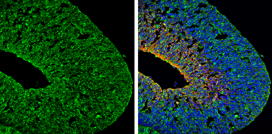
PSD95 antibody detects PSD95 protein expression by immunohistochemical analysis. Sample: Frozen sectioned E13.5 Rat brain. Green: PSD95 protein stained by PSD95 antibody diluted at 1:250. Red: beta Tubulin 3/ TUJ1, a mature neuron marker, stained by beta Tubulin 3/ TUJ1 antibody [GT11710] diluted at 1:500. Blue: Fluoroshield with DAPI .Application
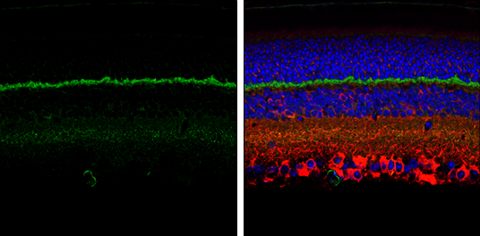
PSD95 antibody detects PSD95 protein expression by immunohistochemical analysis. Sample: Paraffin-Embedded adult mouse retina. Green: PSD95 protein stained by PSD95 antibody diluted at 1:250. Red: beta Tubulin 3/ TUJ1, stained by beta Tubulin 3/ TUJ1 antibody [GT11710] diluted at 1:250. Blue: Fluoroshield with DAPI .Application
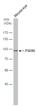
Mouse tissue extract (50 µg) was separated by 7.5% SDS-PAGE, and the membrane was blotted with PSD95 antibody diluted at 1:1000.Application
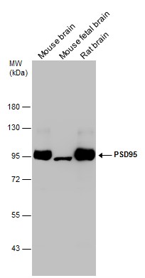
Various tissue extracts (50 µg) were separated by 7.5% SDS-PAGE, and the membrane was blotted with PSD95 antibody diluted at 1:1000.WB
B WB analysis showed that CIH exposure significantly downregulated the expression of NR1 in both WT and APP/PS1 mice. We observed lower expression of PSD95 in WT-CIH, APP/PS1-Sham, and APP/PS1-CIH mice than in WT-Sham mice. The lowest expression of NR1 and PSD95 was observed in APP/PS1-CIH mice. N = 3 mice per group.| Product Name | PSD95 antibody |
|---|---|
| Antibody Type | Primary Antibodies |
| Product description | Rabbit Polyclonal antibody to PSD95 (discs, large homolog 4 (Drosophila))Interacts with the cytoplasmic tail of NMDA receptor subunits and shaker-type potassium channels. Required for synaptic plasticity associated with NMDA receptor signaling. Overexpression or depletion of DLG4 changes the ratio of excitatory to inhibitory synapses in hippocampal neurons. May reduce the amplitude of ACCN3 acid-evoked currents by retaining the channel intracellularly. May regulate the intracellular trafficking of ADR1B. |
| Immunogen | Carrier-protein conjugated synthetic peptide encompassing a sequence within the N-terminus region of mouse PSD95. The exact sequence is proprietary. |
| Clonality | Polyclonal |
|---|---|
| Isotype | IgG |
| Host Species | Rabbit |
| Tested Applications | IHC-FWB |
| *Optimal dilutions/concentrations should be determined by the researcher. | |
| Species Reactivity | MouseRat |
| Predicted Reactivity | Human, BovineHuman (100%), Bovine (100%) |
| Concentration | 1mg/ml |
| Purification | Affinity purified |
| Gene Synonyms | Dlg4 antibody , Dlgh4 antibody , PSD95 antibody , PSD95 antibody , SAP90 antibody , SAP90A antibody , discs, large homolog 4 (Drosophila) antibody |
|---|---|
| Target | PSD95 |
| Molecular Weight(MW) | 80 kDa (note) (2) |
| Cellular Localization | Membrane , Cell junction , synapse , postsynaptic cell membrane , postsynaptic density , synapse |
Application
PSD95 antibody detects PSD95 protein expression by immunohistochemical analysis. Sample: Frozen sectioned E13.5 Rat brain. Green: PSD95 protein stained by PSD95 antibody diluted at 1:250. Red: beta Tubulin 3/ TUJ1, a mature neuron marker, stained by beta Tubulin 3/ TUJ1 antibody [GT11710] diluted at 1:500. Blue: Fluoroshield with DAPI .
Application
PSD95 antibody detects PSD95 protein expression by immunohistochemical analysis. Sample: Paraffin-Embedded adult mouse retina. Green: PSD95 protein stained by PSD95 antibody diluted at 1:250. Red: beta Tubulin 3/ TUJ1, stained by beta Tubulin 3/ TUJ1 antibody [GT11710] diluted at 1:250. Blue: Fluoroshield with DAPI .
Application
Mouse tissue extract (50 µg) was separated by 7.5% SDS-PAGE, and the membrane was blotted with PSD95 antibody diluted at 1:1000.
Application
Various tissue extracts (50 µg) were separated by 7.5% SDS-PAGE, and the membrane was blotted with PSD95 antibody diluted at 1:1000.
WB
B WB analysis showed that CIH exposure significantly downregulated the expression of NR1 in both WT and APP/PS1 mice. We observed lower expression of PSD95 in WT-CIH, APP/PS1-Sham, and APP/PS1-CIH mice than in WT-Sham mice. The lowest expression of NR1 and PSD95 was observed in APP/PS1-CIH mice. N = 3 mice per group.| Positive Control | mouse eye , mouse brain│mouse fetal brain│rat brain |
|---|---|
| Application Notes | *Optimal dilutions/concentrations should be determined by the researcher. |
| Form | Liquid |
|---|---|
| Storage Instructions | Keep as concentrated solution. Aliquot and store at -20ºC or below. Avoid multiple freeze-thaw cycles. |
| Storage Buffer | 1XPBS, 20% Glycerol (pH7). 0.025% ProClin 300 was added as a preservative. |


