Application
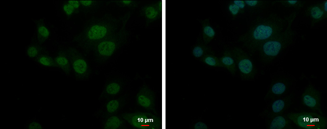
PSMD10 antibody [N1C3] detects PSMD10 protein at cytoplasm and nucleus by immunofluorescent analysis. Sample: HepG2 cells were fixed in 4% paraformaldehyde at RT for 15 min. Green: PSMD10 protein stained by PSMD10 antibody [N1C3] diluted at 1:500. Blue: Hoechst 33342 staining. Scale bar = 10 μm.Application
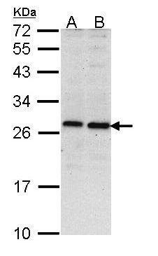
Sample (30 ug of whole cell lysate) A: Hela B: Hep G2 12% SDS PAGE diluted at 1:1000| Product Name | PSMD10 antibody |
|---|---|
| Antibody Type | Primary Antibodies |
| Product description | Rabbit polyclonal antibody to PSMD10 (proteasome (prosome, macropain) 26S subunit, non-ATPase, 10)The 26S proteasome is a multicatalytic proteinase complex with a highly ordered structure composed of 2 complexes, a 20S core and a 19S regulator. The 20S core is composed of 4 rings of 28 non-identical subunits; 2 rings are composed of 7 alpha subunits and 2 rings are composed of 7 beta subunits. The 19S regulator is composed of a base, which contains 6 ATPase subunits and 2 non-ATPase subunits, and a lid, which contains up to 10 non-ATPase subunits. Proteasomes are distributed throughout eukaryotic cells at a high concentration and cleave peptides in an ATP/ubiquitin-dependent process in a non-lysosomal pathway. An essential function of a modified proteasome, the immunoproteasome, is the processing of class I MHC peptides. This gene encodes a non-ATPase subunit of the 19S regulator. Two transcripts encoding different isoforms have been described. Pseudogenes have been identified on chromosomes 3 and 20. [provided by RefSeq] |
| Immunogen | Recombinant protein encompassing a sequence within the center region of human PSMD10. The exact sequence is proprietary. |
| Clonality | Polyclonal |
|---|---|
| Isotype | IgG |
| Host Species | Rabbit |
| Tested Applications | ICC/IFWB |
| *Optimal dilutions/concentrations should be determined by the researcher. | |
| Species Reactivity | Human |
| Predicted Reactivity | Mouse, Rat, Chicken, Rhesus MonkeyMouse (95%), Rat (94%), Chicken (81%), Rhesus Monkey (100%) |
| Concentration | 0.2mg/ml |
| Purification | Affinity purified |
| Gene Synonyms | dJ889N15.2 antibody, p28 antibody, p28(GANK) antibody, PSMD10 antibody, hepatocellular carcinoma-associated protein p28-II antibody, 26S proteasome regulatory subunit p28 antibody, ankyrin repeat protein antibody, gankyrin antibody, 26S proteasome non-ATPa |
|---|---|
| Target | PSMD10 |
| Molecular Weight(MW) | 24 kDa (note) (2) |
Application
PSMD10 antibody [N1C3] detects PSMD10 protein at cytoplasm and nucleus by immunofluorescent analysis. Sample: HepG2 cells were fixed in 4% paraformaldehyde at RT for 15 min. Green: PSMD10 protein stained by PSMD10 antibody [N1C3] diluted at 1:500. Blue: Hoechst 33342 staining. Scale bar = 10 μm.
Application
Sample (30 ug of whole cell lysate) A: Hela B: Hep G2 12% SDS PAGE diluted at 1:1000| Application Notes | *Optimal dilutions/concentrations should be determined by the researcher. |
|---|
| Form | Liquid |
|---|---|
| Storage Instructions | Keep as concentrated solution. Aliquot and store at -20oC or below. Avoid multiple freeze-thaw cycles. |
| Storage Buffer | 0.1M Tris, 0.1M Glycine, 20% Glycerol (pH7). 0.01% Thimerosal was added as a preservative. |


