Application
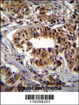
CAD Antibody immunohistochemistry analysis in formalin fixed and paraffin embedded human breast carcinoma followed by peroxidase conjugation of the secondary antibody and DAB staining.Application
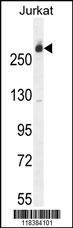
Western blot analysis in Jurkat cell line lysates (35ug/lane).Application
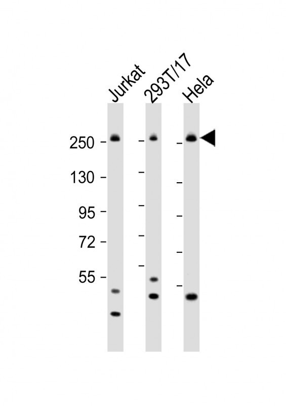
Western Blot at 1:2000 dilution Lane 1: Jurkat whole cell lysate Lane 2: 293T/17 whole cell lysate Lane 3: Hela whole cell lysate Lysates/proteins at 20 ug per lane.Application
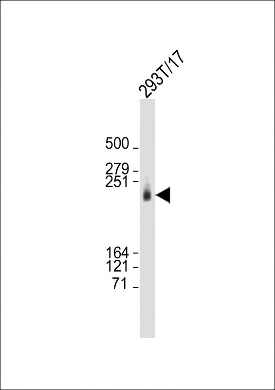
Western Blot at 1:2000 dilution + 293T/17 whole cell lysate Lysates/proteins at 20 ug per lane.Application
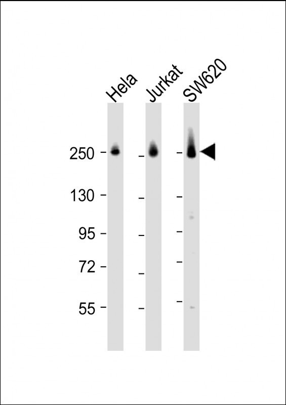
Western Blot at 1:2000 dilution Lane 1: Hela whole cell lysate Lane 2: Jurkat whole cell lysate Lane 3: SW620 whole cell lysate Lysates/proteins at 20 ug per lane.Application
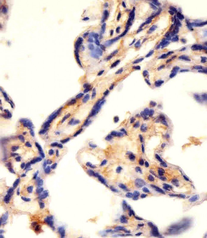
Antibody staining CAD in human placenta tissue sections by Immunohistochemistry (IHC-P - paraformaldehyde-fixed, paraffin-embedded sections).Application
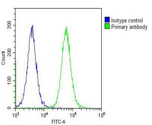
Overlay histogram showing Hela cells stained with Antibody (green line). The cells were fixed with 2% paraformaldehyde (10 min) and then permeabilized with 90% methanol for 10 min. The cells were then icubated in 2% bovine serum albumin to block non-specific protein-protein interactions followed by the antibody (1:25 dilution) for 60 min at 37ºC. The secondary antibody used was Goat-Anti-Rabbit Ig| Product Name | CAD Antibody |
|---|---|
| Antibody Type | Primary Antibodies |
| Product description | The de novo synthesis of pyrimidine nucleotides is required for mammalian cells to proliferate. This gene encodes a trifunctional protein which is associated with the enzymatic activities of the first 3 enzymes in the 6-step pathway of pyrimidine biosynthesis: carbamoylphosphate synthetase (CPS II), aspartate transcarbamoylase, and dihydroorotase. This protein is regulated by the mitogen-activated protein kinase (MAPK) cascade, which indicates a direct link between activation of the MAPK cascade and de novo biosynthesis of pyrimidine nucleotides.1) Jia, P., et al. Schizophr. Res. 122 (1-3), 38-42 (2010) : |
| Immunogen | This CAD antibody is generated from rabbits immunized with a KLH conjugated synthetic peptide between 780-809 amino acids from the Central region of human CAD. |
| Clonality | Polyclonal |
|---|---|
| Isotype | Ig |
| Host Species | Rabbit |
| Tested Applications | FACSIHC-PWB |
| For FACS starting dilution is: 1:25 For IHC-P starting dilution is: 1:25 For WB starting dilution is: 1:2000: |
|
| Species Reactivity | Human |
| Concentration | 1mg/ml |
| Purification | Affinity purified |
| Gene Symbol | CAD |
|---|---|
| Alternative Names | CAD protein Glutamine-dependent carbamoyl-phosphate synthase Aspartate carbamoyltransferase Dihydroorotase CAD |
| Molecular Weight(MW) | 243 kDa |
Application
CAD Antibody immunohistochemistry analysis in formalin fixed and paraffin embedded human breast carcinoma followed by peroxidase conjugation of the secondary antibody and DAB staining.
Application
Western blot analysis in Jurkat cell line lysates (35ug/lane).
Application
Western Blot at 1:2000 dilution Lane 1: Jurkat whole cell lysate Lane 2: 293T/17 whole cell lysate Lane 3: Hela whole cell lysate Lysates/proteins at 20 ug per lane.
Application
Western Blot at 1:2000 dilution + 293T/17 whole cell lysate Lysates/proteins at 20 ug per lane.
Application
Western Blot at 1:2000 dilution Lane 1: Hela whole cell lysate Lane 2: Jurkat whole cell lysate Lane 3: SW620 whole cell lysate Lysates/proteins at 20 ug per lane.
Application
Antibody staining CAD in human placenta tissue sections by Immunohistochemistry (IHC-P - paraformaldehyde-fixed, paraffin-embedded sections).
Application
Overlay histogram showing Hela cells stained with Antibody (green line). The cells were fixed with 2% paraformaldehyde (10 min) and then permeabilized with 90% methanol for 10 min. The cells were then icubated in 2% bovine serum albumin to block non-specific protein-protein interactions followed by the antibody (1:25 dilution) for 60 min at 37ºC. The secondary antibody used was Goat-Anti-Rabbit Ig| Application Notes | For FACS starting dilution is: 1:25 For IHC-P starting dilution is: 1:25 For WB starting dilution is: 1:2000: |
|---|
| Form | Liquid |
|---|---|
| Storage Instructions | Store at 4˚C for three months and -20˚C, stable for up to one year. As with all antibodies care should be taken to avoid repeated freeze thaw cycles. Antibodies should not be exposed to prolonged high temperatures. |
| Storage Buffer | Supplied in PBS with 0.09% (W/V) sodium azide. |


