Application
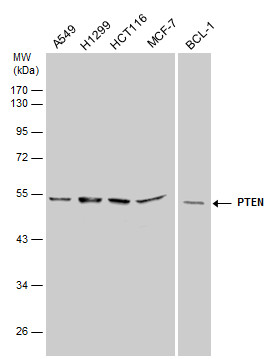
PTEN antibody detects PTEN protein by western blot analysis. Various whole cell extracts (30 µg) were separated by 10% SDS-PAGE, and the membrane was blotted with PTEN antibody diluted at a dilution of 1:500. The HRP-conjugated anti-rabbit IgG antibody was used to detect the primary antibody.Application
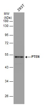
Whole cell extract (30 µg) was separated by 10% SDS-PAGE, and the membrane was blotted with PTEN antibody diluted at 1:500. The HRP-conjugated anti-rabbit IgG antibody was used to detect the primary antibody.Application
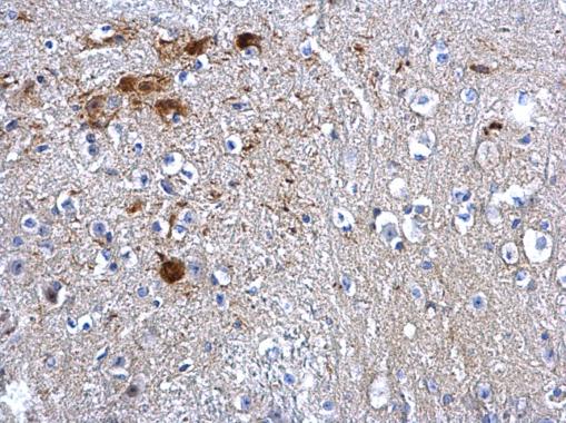
PTEN antibody detects PTEN protein at cytosol and nucleus on mouse middle brain by immunohistochemical analysis. Sample: Paraffin-embedded mouse middle brain. PTEN antibody dilution: 1:500.Application
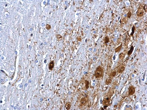
PTEN antibody detects PTEN protein at cytosol and nucleus on rat middle brain by immunohistochemical analysis. Sample: Paraffin-embedded rat middle brain. PTEN antibody dilution: 1:500.Application
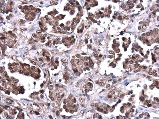
PTEN antibody detects PTEN protein at cytoplasm in human ovarian cancer by immunohistochemical analysis. Sample: Paraffin-embedded human ovarian cancer. PTEN antibody diluted at 1:500.Application
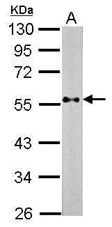
Sample (30 µg of whole cell lysate) A: NIH-3T3 10% SDS PAGE diluted at 1:500 The HRP-conjugated anti-rabbit IgG antibody was used to detect the primary antibody.Application
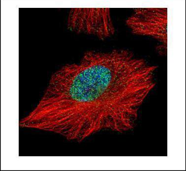
Confocal immunofluorescence analysis (Olympus FV10i) of paraformaldehyde-fixed HeLa, using PTEN antibody (Green) at 1:500 dilution. Alpha-tubulin filaments were labeled with (Red) at 1:2000.| Product Name | PTEN antibody |
|---|---|
| Antibody Type | Primary Antibodies |
| Product description | Rabbit Polyclonal antibody to PTEN (phosphatase and tensin homolog)This gene was identified as a tumor suppressor that is mutated in a large number of cancers at high frequency. The protein encoded this gene is a phosphatidylinositol-3,4,5-trisphosphate 3-phosphatase. It contains a tensin like domain as well as a catalytic domain similar to that of the dual specificity protein tyrosine phosphatases. Unlike most of the protein tyrosine phosphatases, this protein preferentially dephosphorylates phosphoinositide substrates. It negatively regulates intracellular levels of phosphatidylinositol-3,4,5-trisphosphate in cells and functions as a tumor suppressor by negatively regulating AKT/PKB signaling pathway. [provided by RefSeq] |
| Immunogen | Recombinant protein encompassing a sequence within the center region of human PTEN. The exact sequence is proprietary. |
| Clonality | Polyclonal |
|---|---|
| Isotype | IgG |
| Host Species | Rabbit |
| Tested Applications | ICC/IFIHC-PWB |
| *Optimal dilutions/concentrations should be determined by the researcher.
: |
|
| Species Reactivity | HumanMouseRat |
| Concentration | 1mg/ml (Please refer to the vial label for the specific concentration) |
| Purification | Affinity purified |
| Gene Synonyms | 10q23del antibody BZS antibody DEC antibody GLM2 antibody MGC11227 antibody MHAM antibody MMAC1 antibody PTEN1 antibody TEP1 antibody PTEN antibody mutated in multiple advanced cancers 1 antibody phosphatase and tensin-like protein antibody pho |
|---|---|
| Molecular Weight(MW) | 47 kDa (note) (3) |
Application
PTEN antibody detects PTEN protein by western blot analysis. Various whole cell extracts (30 µg) were separated by 10% SDS-PAGE, and the membrane was blotted with PTEN antibody diluted at a dilution of 1:500. The HRP-conjugated anti-rabbit IgG antibody was used to detect the primary antibody.
Application
Whole cell extract (30 µg) was separated by 10% SDS-PAGE, and the membrane was blotted with PTEN antibody diluted at 1:500. The HRP-conjugated anti-rabbit IgG antibody was used to detect the primary antibody.
Application
PTEN antibody detects PTEN protein at cytosol and nucleus on mouse middle brain by immunohistochemical analysis. Sample: Paraffin-embedded mouse middle brain. PTEN antibody dilution: 1:500.
Application
PTEN antibody detects PTEN protein at cytosol and nucleus on rat middle brain by immunohistochemical analysis. Sample: Paraffin-embedded rat middle brain. PTEN antibody dilution: 1:500.
Application
PTEN antibody detects PTEN protein at cytoplasm in human ovarian cancer by immunohistochemical analysis. Sample: Paraffin-embedded human ovarian cancer. PTEN antibody diluted at 1:500.
Application
Sample (30 µg of whole cell lysate) A: NIH-3T3 10% SDS PAGE diluted at 1:500 The HRP-conjugated anti-rabbit IgG antibody was used to detect the primary antibody.
Application
Confocal immunofluorescence analysis (Olympus FV10i) of paraformaldehyde-fixed HeLa, using PTEN antibody (Green) at 1:500 dilution. Alpha-tubulin filaments were labeled with (Red) at 1:2000.| Application Notes | *Optimal dilutions/concentrations should be determined by the researcher.
: |
|---|
| Form | Liquid |
|---|---|
| Storage Instructions | Keep as concentrated solution. Aliquot and store at -20ºC or below. Avoid multiple freeze-thaw cycles. |
| Storage Buffer | 1XPBS, 1% BSA, 20% Glycerol (pH7). 0.01% Thimerosal was added as a preservative. |


