Application
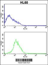
Flow cytometric analysis of HL60 cells (bottom histogram) compared to a negative control cell (top histogram).FITC-conjugated goat-anti-rabbit secondary antibodies were used for the analysis.Application
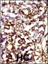
Formalin-fixed and paraffin-embedded human cancer tissue reacted with the primary antibody, which was peroxidase-conjugated to the secondary antibody, followed by AEC staining. BC = breast carcinoma; HC = hepatocarcinoma.Application
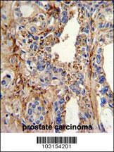
Formalin-fixed and paraffin-embedded human prostate carcinoma with CDK4 Antibody , which was peroxidase-conjugated to the secondary antibody, followed by DAB staining.Application
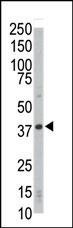
Western blot analysis of anti-CDK4 Pab in HL-60 cell lysateApplication
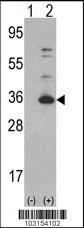
Western blot analysis of CDK4 using rabbit polyclonal CDK4 Antibody using 293 cell lysates (2 ug/lane) either nontransfected (Lane 1) or transiently transfected with the CDK4 gene (Lane 2).Application
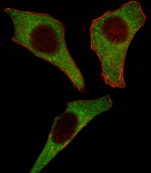
Fluorescent image of Hela cell stained with CDK4 Antibody .Hela cells were fixed with 4% PFA (20 min), permeabilized with Triton X-100 (0.1%, 10 min), then incubated with CDK4 primary antibody (1:25). For secondary antibody, Alexa Fluor 488 conjugated donkey anti-rabbit antibody (green) was used (1:400).Cytoplasmic actin was counterstained with Alexa Fluor 555 (red) conjugated Phalloidin (7units/m| Product Name | CDK4 Antibody |
|---|---|
| Antibody Type | Primary Antibodies |
| Antigen Alias | Cyclin-dependent kinase 4, Cell division protein kinase 4, PSK-J3, CDK4 |
| Product description | CDK4 is a member of the Ser/Thr protein kinase family. This protein is highly similar to the gene products of S. cerevisiae cdc28 and S. pombe cdc2. It is a catalytic subunit of the protein kinase complex that is important for cell cycle G1 phase progression. The activity of this kinase is restricted to the G1-S phase, which is controlled by the regulatory subunits D-type cyclins and CDK inhibitor p16(INK4a). This kinase was shown to be responsible for the phosphorylation of retinoblastoma gene product (Rb). Mutations in this gene as well as in its related proteins including D-type cyclins, p16(INK4a) and Rb were all found to be associated with tumorigenesis of a variety of cancers. Multiple polyadenylation sites of the gene have been reported.1) Mori, N., et al., Int. J. Hematol. 77(3):259-262 (2003). |
| Immunogen | This CDK4 antibody is generated from rabbits immunized with a KLH conjugated synthetic peptide between 273-305 amino acids from the C-terminal region of human CDK4. |
| Clonality | Polyclonal |
|---|---|
| Isotype | Ig |
| Host Species | Rabbit |
| Tested Applications | FACSIFIHC-PWB |
| For IF starting dilution is: 1:10~50 For WB starting dilution is: 1:1000 For IHC-P starting dilution is: 1:50~100 For FACS starting dilution is: 1:10~50: |
|
| Species Reactivity | Human |
| Concentration | 1mg/ml |
| Gene Symbol | CDK4 |
|---|---|
| Alternative Names | Cyclin-dependent kinase 4 Cell division protein kinase 4 PSK-J3 CDK4 |
| Molecular Weight(MW) | 34 kDa |
Application
Flow cytometric analysis of HL60 cells (bottom histogram) compared to a negative control cell (top histogram).FITC-conjugated goat-anti-rabbit secondary antibodies were used for the analysis.
Application
Formalin-fixed and paraffin-embedded human cancer tissue reacted with the primary antibody, which was peroxidase-conjugated to the secondary antibody, followed by AEC staining. BC = breast carcinoma; HC = hepatocarcinoma.
Application
Formalin-fixed and paraffin-embedded human prostate carcinoma with CDK4 Antibody , which was peroxidase-conjugated to the secondary antibody, followed by DAB staining.
Application
Western blot analysis of anti-CDK4 Pab in HL-60 cell lysate
Application
Western blot analysis of CDK4 using rabbit polyclonal CDK4 Antibody using 293 cell lysates (2 ug/lane) either nontransfected (Lane 1) or transiently transfected with the CDK4 gene (Lane 2).
Application
Fluorescent image of Hela cell stained with CDK4 Antibody .Hela cells were fixed with 4% PFA (20 min), permeabilized with Triton X-100 (0.1%, 10 min), then incubated with CDK4 primary antibody (1:25). For secondary antibody, Alexa Fluor 488 conjugated donkey anti-rabbit antibody (green) was used (1:400).Cytoplasmic actin was counterstained with Alexa Fluor 555 (red) conjugated Phalloidin (7units/m| Application Notes | For IF starting dilution is: 1:10~50 For WB starting dilution is: 1:1000 For IHC-P starting dilution is: 1:50~100 For FACS starting dilution is: 1:10~50: |
|---|
| Form | Liquid |
|---|---|
| Storage Instructions | Store at 4˚C for three months and -20˚C, stable for up to one year. As with all antibodies care should be taken to avoid repeated freeze thaw cycles. Antibodies should not be exposed to prolonged high temperatures. |
| Storage Buffer | Supplied in PBS with 0.09% (W/V) sodium azide. |


