Application
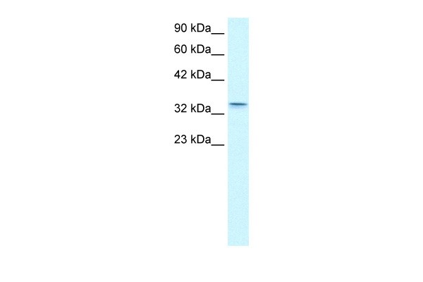
Antibody used in WB on Human kidney at 0.2-1 ug/ml.| Product Name | CLDN16 Antibody |
|---|---|
| Antibody Type | Primary Antibodies |
| Antigen Alias | CLDN16, HOMG3, PCLN1 |
| Product description | Tight junctions represent one mode of cell-to-cell adhesion in epithelial or endothelial cell sheets, forming continuous seals around cells and serving as a physical barrier to prevent solutes and water from passing freely through the paracellular space. These junctions are comprised of sets of continuous networking strands in the outwardly facing cytoplasmic leaflet, with complementary grooves in the inwardly facing extracytoplasmic leaflet. Claudin-16, a member of the claudin family, is an integral membrane protein and a component of tight junction strands. It is found primarily in the kidneys, specifically in the thick ascending limb of Henle, where it acts as either an intercellular pore or ion concentration sensor to regulate the paracellular resorption of magnesium ions. Defects in the corresponding gene are a cause of primary hypomagnesemia, which is characterized by massive renal magnesium wasting with hypomagnesemia and hypercalciuria, resulting in nephrocalcinosis and renal failure.Tight junctions represent one mode of cell-to-cell adhesion in epithelial or endothelial cell sheets, forming continuous seals around cells and serving as a physical barrier to prevent solutes and water from passing freely through the paracellular space. These junctions are comprised of sets of continuous networking strands in the outwardly facing cytoplasmic leaflet, with complementary grooves in the inwardly facing extracytoplasmic leaflet. The protein encoded by this gene, a member of the claudin family, is an integral membrane protein and a component of tight junction strands. It is found primarily in the kidneys, specifically in the thick ascending limb of Henle, where it acts as either an intercellular pore or ion concentration sensor to regulate the paracellular resorption of magnesium ions. Defects in this gene are a cause of primary hypomagnesemia, which is characterized by massive renal magnesium wasting with hypomagnesemia and hypercalciuria, resulting in nephrocalcinosis and renal failure. Publication Note: This RefSeq record includes a subset of the publications that are available for this gene. Please see the Entrez Gene record to access additional publications.1) Konrad, M., (2008) J. Am. Soc. Nephrol. 19 (1), 171-181. |
| Immunogen | Antibody produced in rabbits immunized with a synthetic peptide corresponding a region of human CLDN16. |
| Clonality | Polyclonal |
|---|---|
| Host Species | Rabbit |
| Tested Applications | ELISAWB |
| CLDN16 antibody can be used for detection of CLDN16 by ELISA at 1:312500. CLDN16 antibody can be used for detection of CLDN16 by western blot at 1 μg/mL, and HRP conjugated secondary antibody should be diluted 1:50,000 - 100,000.: |
|
| Species Reactivity | Human |
| Concentration | 1mg/ml |
| Purification | Affinity purified |
| Gene Symbol | CLDN16 |
|---|---|
| Alternative Names | CLDN16 HOMG3 PCLN1 |
| Molecular Weight(MW) | 34 kDa |
Application
Antibody used in WB on Human kidney at 0.2-1 ug/ml.| Positive Control | 1) Cat. No. 1211 - HepG2 Cell Lysate |
|---|---|
| Application Notes | CLDN16 antibody can be used for detection of CLDN16 by ELISA at 1:312500. CLDN16 antibody can be used for detection of CLDN16 by western blot at 1 μg/mL, and HRP conjugated secondary antibody should be diluted 1:50,000 - 100,000.: |
| Form | Liquid |
|---|---|
| Storage Instructions | For short periods of storage (days) store at 4˚C. For longer periods of storage, store CLDN16 antibody at -20˚C. As with any antibody avoid repeat freeze-thaw cycles. |
| Storage Buffer | Antibody is lyophilized in PBS buffer with 2% sucrose. Add 50 μL of distilled water. Final antibody concentration is 1 mg/mL. |


