ELISA
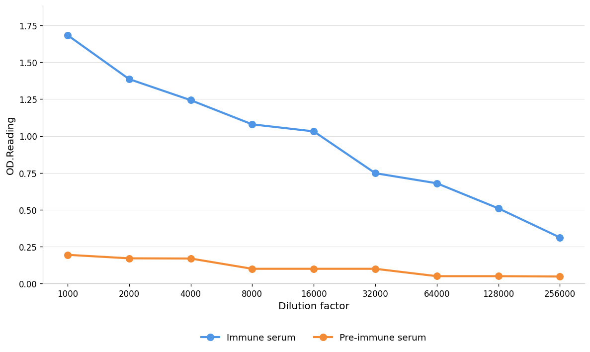
Coat the plate with immunizing antigen.Blue line: Immune serum; Orange line: Pre-immune serum.WB
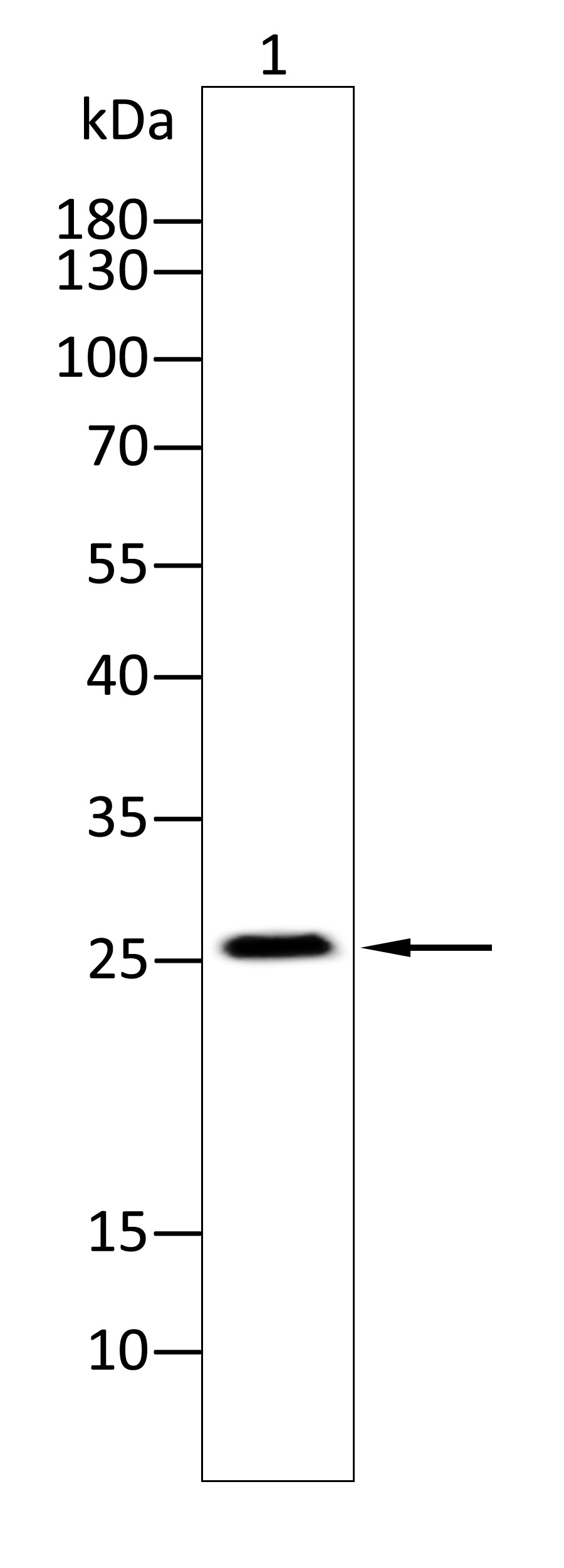
Western blot analysis using CLDN16 antibody against Rat kidney tissue(1) . 12% SDS-PAGE gel.Sample loading: 20μg /lane. Transfer the proteins onto a PVDF membrane (OM790003), and block it with TBST (OM750016) plus skimmed milk powder(OM750084) for one hour. Dilute the primary antibody with the antibody diluent (OM750012) at a ratio of 1:1000, and incubate it overnight at 4°C. Wash the membrane three times with TBST (OM750016), 5 minutes each time. At room temperature, dilute the secondary antibody, Goat Anti-Rabbit IgG(H&L)-HRP (OM643487), at a ratio of 1:20000 and incubate for one hour. Wash the membrane three times with TBST (OM750016) again, 5 minutes each time. Use ECL (OM625701) for luminescence.staining time: 60S.| Product Name | Anti-CLDN16 antibody |
|---|---|
| Antibody Type | Primary Antibodies |
| Immunogen | Polypeptide |
| Clonality | Polyclonal |
|---|---|
| Isotype | IgG |
| Host Species | Rabbit |
| Tested Applications | ELISAWB |
| WB:1:1000-1:2000 ELISA:1:1000-1:10000 |
|
| Species Reactivity | HumanMouseRat |
| Concentration | 1mg/ml |
| Purification | Protein A |
| Gene Symbol | CLDN16 |
|---|---|
| Gene Synonyms | claudin-16 HOMG3 PCLN1 |
| Gene Full Name | claudin 16 |
| Gene Summary | Tight junctions represent one mode of cell-to-cell adhesion in epithelial or endothelial cell sheets, forming continuous seals around cells and serving as a physical barrier to prevent solutes and water from passing freely through the paracellular space. These junctions are comprised of sets of continuous networking strands in the outwardly facing cytoplasmic leaflet, with complementary grooves in the inwardly facing extracytoplasmic leaflet. The protein encoded by this gene, a member of the claudin family, is an integral membrane protein and a component of tight junction strands. It is found primarily in the kidneys, specifically in the thick ascending limb of Henle, where it acts as either an intercellular pore or ion concentration sensor to regulate the paracellular resorption of magnesium ions. Defects in this gene are a cause of primary hypomagnesemia, which is characterized by massive renal magnesium wasting with hypomagnesemia and hypercalciuria, resulting in nephrocalcinosis and renal failure. This gene and the CLDN1 gene are clustered on chromosome 3q28. [provided by RefSeq, Jun 2010] |
| Alternative Names | H59D2a protein hypomagnesemia 3 with hypercalciuria and nephrocalcinosis paracellin-1 PCLN-1 renal tight junction protein |
| Molecular Weight(MW) | 26kDa |
| Source | Rabbit |
| Cellular Localization | Cell junction, tight junction;Cell membrane |
| SwissProt ID | Q9Y5I7 |
|---|
ELISA
Coat the plate with immunizing antigen.Blue line: Immune serum; Orange line: Pre-immune serum.
WB
Western blot analysis using CLDN16 antibody against Rat kidney tissue(1) . 12% SDS-PAGE gel.Sample loading: 20μg /lane. Transfer the proteins onto a PVDF membrane (OM790003), and block it with TBST (OM750016) plus skimmed milk powder(OM750084) for one hour. Dilute the primary antibody with the antibody diluent (OM750012) at a ratio of 1:1000, and incubate it overnight at 4°C. Wash the membrane three times with TBST (OM750016), 5 minutes each time. At room temperature, dilute the secondary antibody, Goat Anti-Rabbit IgG(H&L)-HRP (OM643487), at a ratio of 1:20000 and incubate for one hour. Wash the membrane three times with TBST (OM750016) again, 5 minutes each time. Use ECL (OM625701) for luminescence.staining time: 60S.| Application Notes | WB:1:1000-1:2000 ELISA:1:1000-1:10000 |
|---|
| Form | Liquid |
|---|---|
| Storage Instructions | Shipped at 4°C. Store at +4°C short term (1-2 weeks). Store at -20°C long term. Avoid freeze / thaw cycle. |
| Storage Buffer | Purified antibody in PBS with 0.05% PC300,40% Glycerol. |


