Application
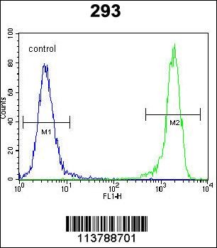
Flow cytometric analysis of 293 cells (right histogram) compared to a negative control cell (left histogram).FITC-conjugated goat-anti-rabbit secondary antibodies were used for the analysis.Application
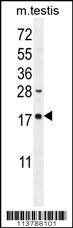
Western blot analysis in mouse testis tissue lysates (35ug/lane).This demonstrates the DDIT3 antibody detected the DDIT3 protein (arrow).Application
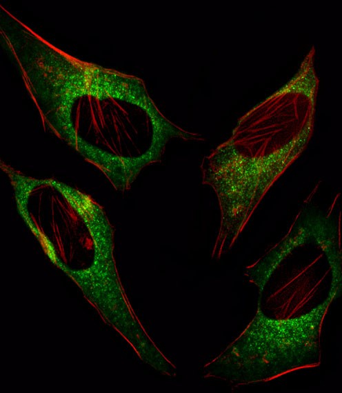
Fluorescent image of Hela cell stained with .Hela cells were fixed with 4% PFA (20 min), permeabilized with Triton X-100 (0.1%, 10 min), then incubated with DDIT3 primary antibody (1:25). For secondary antibody, Alexa Fluor 488 conjugated donkey anti-rabbit antibody (green) was used (1:400).Cytoplasmic actin was counterstained with Alexa Fluor 555 (red) conjugated Phalloidin (7units/ml). DDIT3 immApplication
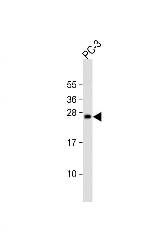
Western Blot at 1:2000 dilution + PC-3 whole cell lysate Lysates/proteins at 20 ug per lane.Application
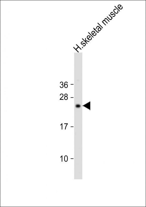
Western Blot at 1:2000 dilution + human skeletal muscle lysate Lysates/proteins at 20 ug per lane.Application
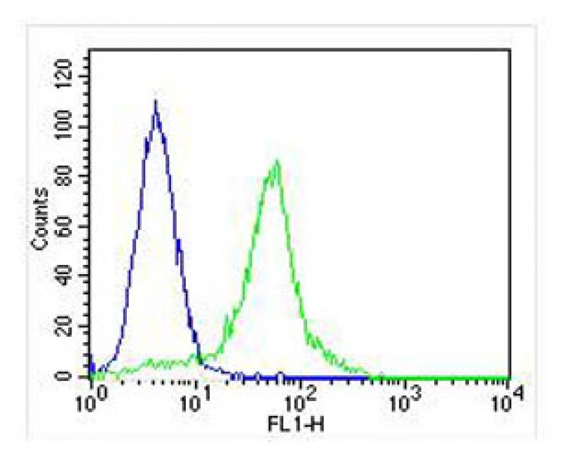
Overlay histogram showing Hela cells stained with Antibody (green line). The cells were fixed with 2% paraformaldehyde (10 min) and then permeabilized with 90% methanol for 10 min. The cells were then icubated in 2% bovine serum albumin to block non-specific protein-protein interactions followed by the antibody (1:25 dilution) for 60 min at 37ºC. The secondary antibody used was Goat-Anti-Rabbit IgApplication
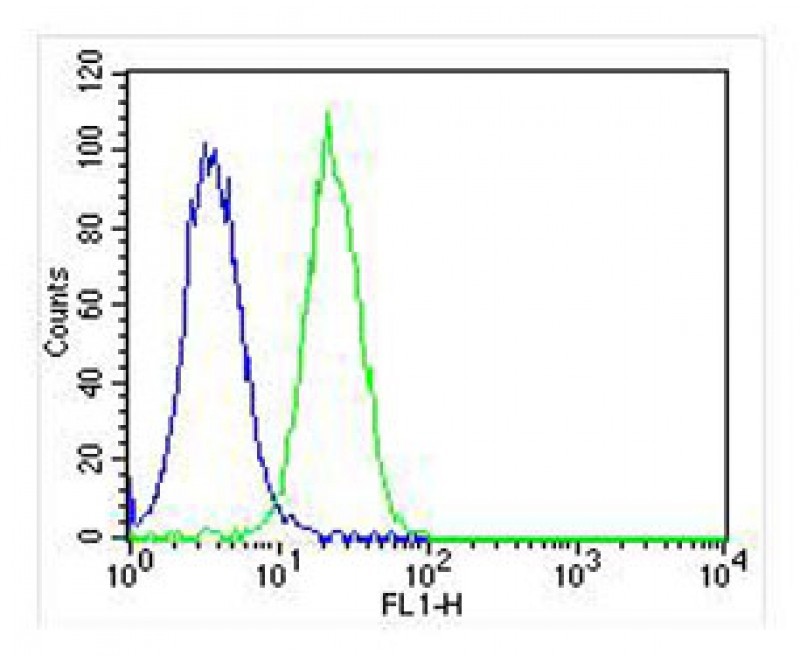
Overlay histogram showing Hela cells stained with Antibody (green line). The cells were fixed with 2% paraformaldehyde (10 min) and then permeabilized with 90% methanol for 10 min. The cells were then icubated in 2% bovine serum albumin to block non-specific protein-protein interactions followed by the antibody (1:25 dilution) for 60 min at 37ºC. The secondary antibody used was Goat-Anti-Rabbit Ig| Product Name | DDIT3 Antibody |
|---|---|
| Antibody Type | Primary Antibodies |
| Product description | This gene encodes a member of the CCAAT/enhancer-binding protein (C/EBP) family of transcription factors. The protein functions as a dominant-negative inhibitor by forming heterodimers with other C/EBP members, such as C/EBP and LAP (liver activator protein), and preventing their DNA binding activity. The protein is implicated in adipogenesis and erythropoiesis, is activated by endoplasmic reticulum stress, and promotes apoptosis. Fusion of this gene and FUS on chromosome 16 or EWSR1 on chromosome 22 induced by translocation generates chimeric proteins in myxoid liposarcomas or Ewing sarcoma. Multiple alternatively spliced transcript variants encoding two isoforms with different length have been identified.1) Park, S.H., et al. J. Immunol. 185(9):5522-5530(2010) |
| Immunogen | This DDIT3 antibody is generated from rabbits immunized with a KLH conjugated synthetic peptide between 120-149 amino acids from the C-terminal region of human DDIT3. |
| Clonality | Polyclonal |
|---|---|
| Isotype | Ig |
| Host Species | Rabbit |
| Tested Applications | ELISAFACSIFIHCWB |
| For FACS starting dilution is: 1:25 For WB starting dilution is: 1:2000 For IF starting dilution is: 1:10~50 | |
| Species Reactivity | HumanMonkeyMouseRat |
| Concentration | 1mg/ml |
| Purification | Affinity purified |
| Gene Symbol | DDIT3 |
|---|---|
| Alternative Names | DNA damage-inducible transcript 3 protein DDIT-3 C/EBP zeta C/EBP-homologous protein CHOP C/EBP-homologous protein 10 CHOP-10 CCAAT/enhancer-binding protein homologous protein Growth arrest and DNA damage-inducible protein GADD153 DDIT3 CHOP CHO |
| Molecular Weight(MW) | 19 kDa |
| Sequence Similarities | Predicted species reactivity based on immunogen sequence: Hamster |
Application
Flow cytometric analysis of 293 cells (right histogram) compared to a negative control cell (left histogram).FITC-conjugated goat-anti-rabbit secondary antibodies were used for the analysis.
Application
Western blot analysis in mouse testis tissue lysates (35ug/lane).This demonstrates the DDIT3 antibody detected the DDIT3 protein (arrow).
Application
Fluorescent image of Hela cell stained with .Hela cells were fixed with 4% PFA (20 min), permeabilized with Triton X-100 (0.1%, 10 min), then incubated with DDIT3 primary antibody (1:25). For secondary antibody, Alexa Fluor 488 conjugated donkey anti-rabbit antibody (green) was used (1:400).Cytoplasmic actin was counterstained with Alexa Fluor 555 (red) conjugated Phalloidin (7units/ml). DDIT3 imm
Application
Western Blot at 1:2000 dilution + PC-3 whole cell lysate Lysates/proteins at 20 ug per lane.
Application
Western Blot at 1:2000 dilution + human skeletal muscle lysate Lysates/proteins at 20 ug per lane.
Application
Overlay histogram showing Hela cells stained with Antibody (green line). The cells were fixed with 2% paraformaldehyde (10 min) and then permeabilized with 90% methanol for 10 min. The cells were then icubated in 2% bovine serum albumin to block non-specific protein-protein interactions followed by the antibody (1:25 dilution) for 60 min at 37ºC. The secondary antibody used was Goat-Anti-Rabbit Ig
Application
Overlay histogram showing Hela cells stained with Antibody (green line). The cells were fixed with 2% paraformaldehyde (10 min) and then permeabilized with 90% methanol for 10 min. The cells were then icubated in 2% bovine serum albumin to block non-specific protein-protein interactions followed by the antibody (1:25 dilution) for 60 min at 37ºC. The secondary antibody used was Goat-Anti-Rabbit Ig| Application Notes | For FACS starting dilution is: 1:25 For WB starting dilution is: 1:2000 For IF starting dilution is: 1:10~50 |
|---|
| Form | Liquid |
|---|---|
| Storage Instructions | Store at 4˚C for three months and -20˚C, stable for up to one year. As with all antibodies care should be taken to avoid repeated freeze thaw cycles. Antibodies should not be exposed to prolonged high temperatures. |
| Storage Buffer | Supplied in PBS with 0.09% (W/V) sodium azide. |


