Application
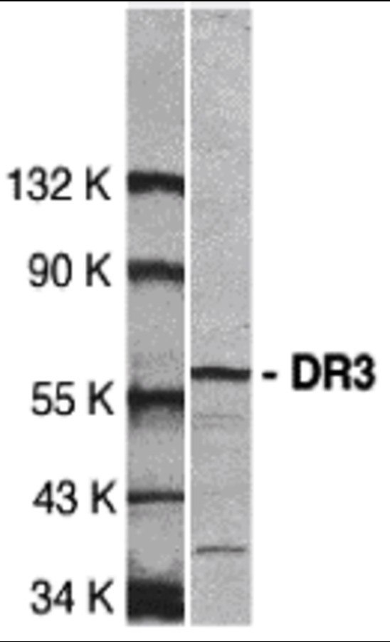
Western blot analysis of DR3 in Jurkat total cell lysate with DR3 antibody at 1:500 dilution.| Product Name | DR3 Antibody |
|---|---|
| Antibody Type | Primary Antibodies |
| Antigen Alias | DR3 Antibody: DR3, TR3, DDR3, LARD, APO-3, TRAMP, WSL-1, WSL-LR, TNFRSF12, APO3, DR3, WSL, WSL1, UNQ455/PRO779, Tumor necrosis factor receptor superfamily member 25, Apo-3 |
| Product description | DR3 Antibody: Apoptosis, or programmed cell death, occurs during normal cellular differentiation and development of multicellular organisms. Apoptosis is induced by certain cytokines including TNF and Fas ligand of the TNF family through their death domain containing receptors, TNFR1 and Fas. A novel cell death receptor was recently identified by several groups independently and designated DR3, Wsl-1, Apo-3, TRAMP and LARD1-5. The ligand for this novel cell death receptor has not yet been defined. DR3 is highly expressed in the tissues enriched in lymphocytes including PBL, thymus and spleen. Like TNFR1, DR3 induces apoptosis and NF-κB activation.1) Chinnaiyan AM; O'Rourke K; Yu GL; Lyons RH; Garg M; Duan DR; Xing L; Gentz R; Ni J; Dixit VM. Science, 1996;274:990- |
| Immunogen | DR3 antibody was raised against a 20 amino acid peptide near the carboxy terminus of human DR3. The immunogen is located within the last 50 amino acids of DR3. |
| Clonality | Polyclonal |
|---|---|
| Isotype | IgG |
| Host Species | Rabbit |
| Tested Applications | ELISAWB |
| DR3 antibody can be used for Western blot 0.5 μg/mL. A 59 kDa band should be detected.: |
|
| Species Reactivity | HumanMouseRat |
| Concentration | 1mg/ml |
| Purification | Affinity purified |
| Gene Symbol | TNFRSF25 |
|---|---|
| Alternative Names | DR3 Antibody: DR3 TR3 DDR3 LARD APO-3 TRAMP WSL-1 WSL-LR TNFRSF12 APO3 DR3 WSL WSL1 UNQ455/PRO779 Tumor necrosis factor receptor superfamily member 25 Apo-3 |
| Molecular Weight(MW) | 59 kDa |
| Tissue Specificity | No cross reaction to other death receptors. |
Application
Western blot analysis of DR3 in Jurkat total cell lysate with DR3 antibody at 1:500 dilution.| Positive Control | 1) Cat. No. 1205 - Jurkat Cell Lysate |
|---|---|
| Application Notes | DR3 antibody can be used for Western blot 0.5 μg/mL. A 59 kDa band should be detected.: |
| Form | Liquid |
|---|---|
| Storage Instructions | DR3 antibody can be stored at 4˚C for three months and -20˚C, stable for up to one year. As with all antibodies care should be taken to avoid repeated freeze thaw cycles. Antibodies should not be exposed to prolonged high temperatures. |
| Storage Buffer | DR3 Antibody is supplied in PBS containing 0.02% sodium azide. |


