WB
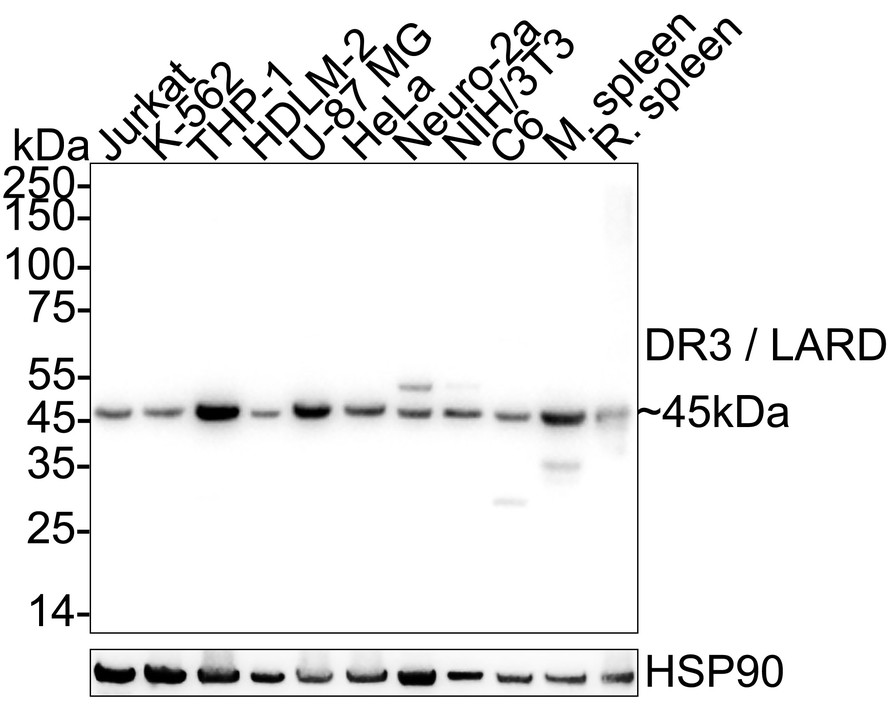
Western blot analysis of DR3 / LARD on different lysates with Rabbit anti-DR3 / LARD antibody at 1/5000 dilution. Lane 1: Jurkat cell lysate (20 µg/Lane), Lane 2: K-562 cell lysate (20 µg/Lane), Lane 3: THP-1 cell lysate (20 µg/Lane), Lane 4: HDLM-2 cell lysate (20 µg/Lane), Lane 5: U-87 MG cell lysate (20 µg/Lane), Lane 6: HeLa cell lysate (20 µg/Lane), Lane 7: Neuro-2a cell lysate (20 µg/Lane), Lane 8: NIH/3T3 cell lysate (20 µg/Lane), Lane 9: C6 cell lysate (20 µg/Lane), Lane 10: Mouse spleen tissue lysate (40 µg/Lane), Lane 11: Rat spleen tissue lysate (40 µg/Lane), Exposure time: 10 seconds; 4-20% SDS-PAGE gel. Proteins were transferred to a PVDF membrane and blocked with 5% NFDM/TBST for 1 hour at room temperature. The primary antibody at 1/5,000 dilution was used in primary antibody dilution at 4℃ overnight. Goat Anti-Rabbit IgG - HRP Secondary Antibody at 1/50,000 dilution was used for 1 hour at room temperature.ICC/IF
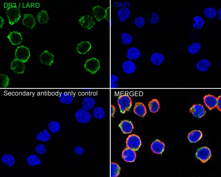
Immunocytochemistry analysis of Jurkat cells labeling DR3 / LARD with Rabbit anti-DR3 / LARD antibody at 1/100 dilution. Cells were fixed in 4% paraformaldehyde for 15 minutes at room temperature, permeabilized with 0.1% Triton X-100 in PBS for 15 minutes at room temperature, then blocked with 1% BSA in 10% negative goat serum for 1 hour at room temperature. Cells were then incubated with Rabbit anti-DR3 / LARD antibody at 1/100 dilution in 1% BSA in PBST overnight at 4 ℃. Goat Anti-Rabbit IgG H&L (488) was used as the secondary antibody at 1/1,000 dilution. PBS instead of the primary antibody was used as the secondary antibody only control. Nuclear DNA was labelled in blue with DAPI. Beta tubulin ( red) was stained at 1/100 dilution overnight at +4℃. Goat Anti-Mouse IgG H&L (594) was used as the secondary antibody at 1/1000 dilution.| Product Name | DR3 / LARD Recombinant Rabbit Monoclonal Antibody |
|---|---|
| Antibody Type | Primary Antibodies |
| Immunogen | Recombinant protein within human DR3 / LARD aa 1-199. |
| Clonality | Monoclonal |
|---|---|
| Isotype | IgG |
| Host Species | Rabbit |
| Tested Applications | ICC/IFWB |
| WB:1:5000 ICC/IF:1:100 |
|
| Species Reactivity | HumanMouseRat |
| Concentration | 1mg/ml |
| Purification | Affinity purified |
| Gene Symbol | TNFRSF25 |
|---|---|
| Gene Synonyms | DR3 TR3 DDR3 LARD APO-3 TRAMP WSL-1 WSL-LR TNFRSF12 |
| Gene Full Name | TNF receptor superfamily member 25 |
| Gene Summary | The protein encoded by this gene is a member of the TNF-receptor superfamily. This receptor is expressed preferentially in the tissues enriched in lymphocytes, and it may play a role in regulating lymphocyte homeostasis. This receptor has been shown to stimulate NF-kappa B activity and regulate cell apoptosis. The signal transduction of this receptor is mediated by various death domain containing adaptor proteins. Knockout studies in mice suggested the role of this gene in the removal of self-reactive T cells in the thymus. Multiple alternatively spliced transcript variants of this gene encoding distinct isoforms have been reported, most of which are potentially secreted molecules. The alternative splicing of this gene in B and T cells encounters a programmed change upon T-cell activation, which predominantly produces full-length, membrane bound isoforms, and is thought to be involved in controlling lymphocyte proliferation induced by T-cell activation. [provided by RefSeq, Jul 2008] |
| Alternative Names | Apo 3 antibody Apo-3 antibody Apo3 antibody Apoptosis inducing receptor AIR antibody Apoptosis inducing receptor antibody Apoptosis mediating receptor antibody Apoptosis mediating receptor DR 3 antibody Apoptosis mediating receptor DR3 antibody Apoptosis mediating receptor TRAMP antibody Apoptosis-inducing receptor AIR antibody Apo 3 antibody Apo-3 antibody Apo3 antibody Apoptosis inducing receptor AIR antibody Apoptosis inducing receptor antibody Apoptosis mediating receptor antibody Apoptosis mediating receptor DR 3 antibody Apoptosis mediating receptor DR3 antibody Apoptosis mediating receptor TRAMP antibody Apoptosis-inducing receptor AIR antibody Apoptosis-mediating receptor DR3 antibody Apoptosis-mediating receptor TRAMP antibody DDR 3 antibody DDR3 antibody Death domain receptor 3 antibody Death domain receptor 3 soluble form antibody Death receptor 3 antibody Death receptor beta antibody DR 3 antibody DR3 antibody LARD antibody Lymphocyte associated receptor of death antibody Lymphocyte-associated receptor of death antibody Protein WSL antibody Protein WSL-1 antibody TNF receptor superfamily member 25 antibody TNFR25 antibody TNFRSF 12 antibody TNFRSF 25 antibody TNFRSF12 antibody TNFRSF12 formerly antibody TNFRSF25 antibody TNR25_HUMAN antibody TR 3 antibody TR3 antibody TRAMP antibody Translocating chain association membrane protein antibody Tumor necrosis factor receptor superfamily member 12 antibody Tumor necrosis factor receptor superfamily member 25 antibody Tumor necrosis factor receptor superfamily member 12 (translocating chain association membrane protein) antibody Tumor necrosis factor receptor superfamily member 12 formerly antibody WSL 1 antibody WSL antibody WSL LR antibody WSL protein antibody WSL1 antibody WSL1 protein antibody WSLLR antibody |
| Molecular Weight(MW) | 45kDa(Observed band size:45kDa) |
| Cellular Localization | Cell membrane; Secreted. |
WB
Western blot analysis of DR3 / LARD on different lysates with Rabbit anti-DR3 / LARD antibody at 1/5000 dilution. Lane 1: Jurkat cell lysate (20 µg/Lane), Lane 2: K-562 cell lysate (20 µg/Lane), Lane 3: THP-1 cell lysate (20 µg/Lane), Lane 4: HDLM-2 cell lysate (20 µg/Lane), Lane 5: U-87 MG cell lysate (20 µg/Lane), Lane 6: HeLa cell lysate (20 µg/Lane), Lane 7: Neuro-2a cell lysate (20 µg/Lane), Lane 8: NIH/3T3 cell lysate (20 µg/Lane), Lane 9: C6 cell lysate (20 µg/Lane), Lane 10: Mouse spleen tissue lysate (40 µg/Lane), Lane 11: Rat spleen tissue lysate (40 µg/Lane), Exposure time: 10 seconds; 4-20% SDS-PAGE gel. Proteins were transferred to a PVDF membrane and blocked with 5% NFDM/TBST for 1 hour at room temperature. The primary antibody at 1/5,000 dilution was used in primary antibody dilution at 4℃ overnight. Goat Anti-Rabbit IgG - HRP Secondary Antibody at 1/50,000 dilution was used for 1 hour at room temperature.
ICC/IF
Immunocytochemistry analysis of Jurkat cells labeling DR3 / LARD with Rabbit anti-DR3 / LARD antibody at 1/100 dilution. Cells were fixed in 4% paraformaldehyde for 15 minutes at room temperature, permeabilized with 0.1% Triton X-100 in PBS for 15 minutes at room temperature, then blocked with 1% BSA in 10% negative goat serum for 1 hour at room temperature. Cells were then incubated with Rabbit anti-DR3 / LARD antibody at 1/100 dilution in 1% BSA in PBST overnight at 4 ℃. Goat Anti-Rabbit IgG H&L (488) was used as the secondary antibody at 1/1,000 dilution. PBS instead of the primary antibody was used as the secondary antibody only control. Nuclear DNA was labelled in blue with DAPI. Beta tubulin ( red) was stained at 1/100 dilution overnight at +4℃. Goat Anti-Mouse IgG H&L (594) was used as the secondary antibody at 1/1000 dilution.| Application Notes | WB:1:5000 ICC/IF:1:100 |
|---|
| Form | Liquid |
|---|---|
| Storage Instructions | Shipped at 4℃. Store at +4℃ short term (1-2 weeks). It is recommended to aliquot into single-use upon delivery. Store at -20℃ long term. |
| Storage Buffer | PBS (pH7.4), 0.1% BSA, 40% Glycerol. Preservative: 0.05% Sodium Azide. |


