Application
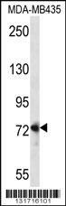
Western blot analysis in MDA-MB435 cell line lysates (35ug/lane).Application
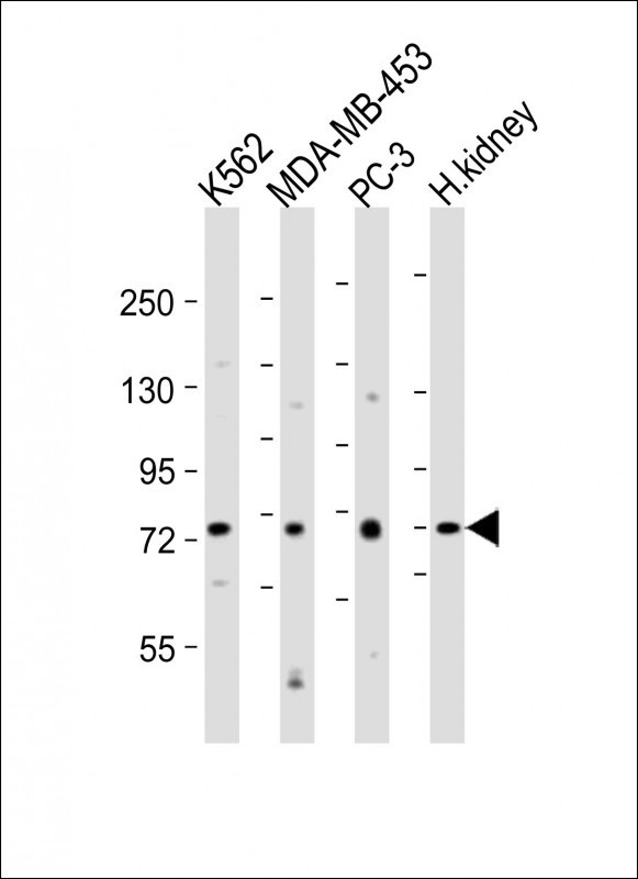
Western Blot at 1:2000 dilution Lane 1: K562 whole cell lysates Lane 2: MDA-MB-453 whole cell lysates Lane 3: PC-3 whole cell lysates Lane 4: human kidney lysates Lysates/proteins at 20 ug per lane.Application
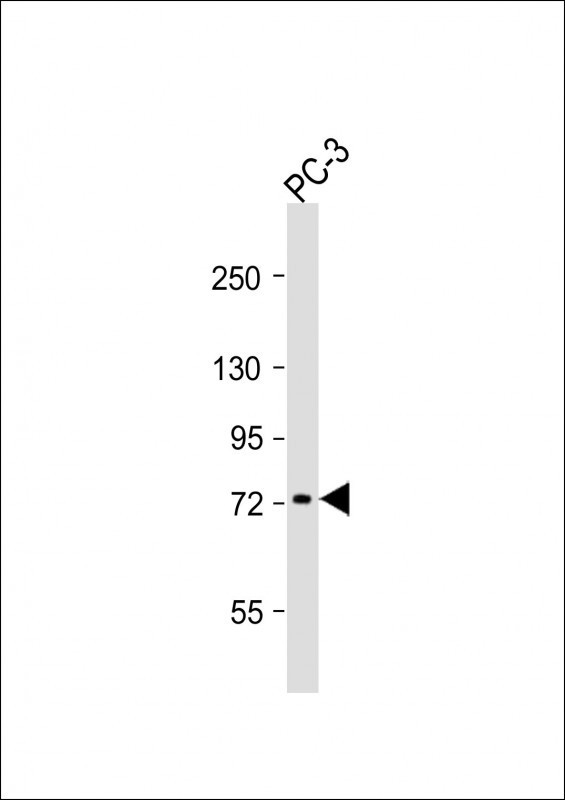
Western Blot at 1:1000 dilution + PC-3 whole cell lysates Lysates/proteins at 20 ug per lane.Application
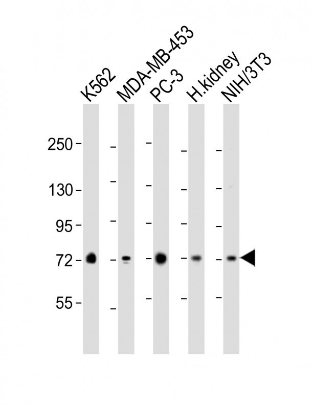
Western Blot at 1:2000 dilution Lane 1: K562 whole cell lysate Lane 2: MDA-MB-453 whole cell lysate Lane 3: PC-3 whole cell lysate Lane 4: human kidney lysate Lane 5: NIH/3T3 whole cell lysate Lysates/proteins at 20 ug per lane.Application
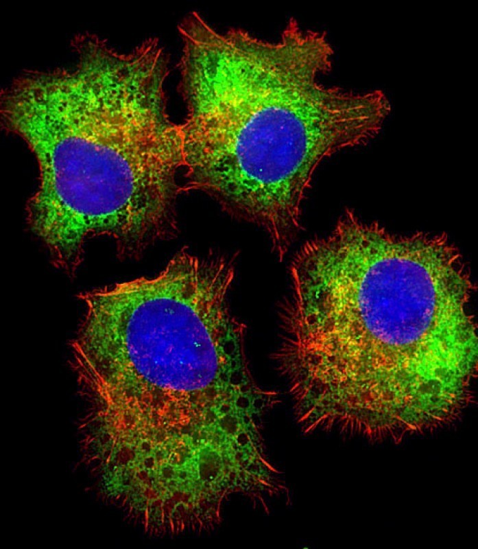
Immunofluorescent analysis of 4% paraformaldehyde-fixed, 0. 1% Triton X-100 permeabilized HepG2 (human liver hepatocellular carcinoma cell line) cells labeling Pdx1 with antibody at 1/25 dilution, followed by Dylight 488-conjugated goat anti-rabbit IgG (NK179883) secondary antibody at 1/200 dilution (green). Immunofluorescence image showing cytoplasm staining on HepG2 cell line. Cytoplasmic actinApplication
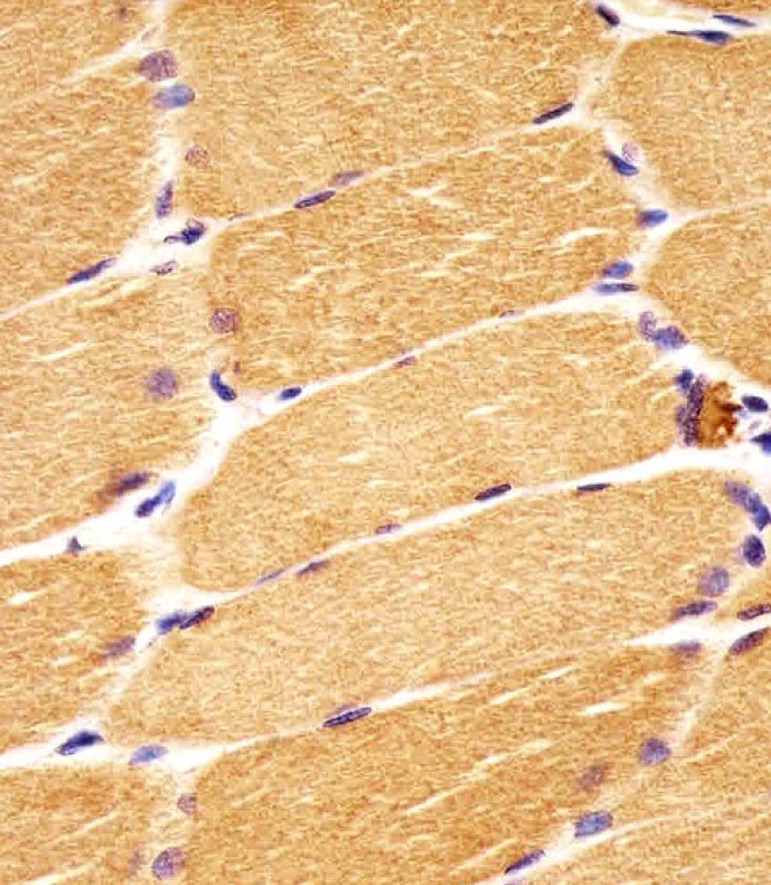
Antibody staining DVL1 in human skeletal muscle sections by Immunohistochemistry (IHC-P - paraformaldehyde-fixed, paraffin-embedded sections).Application
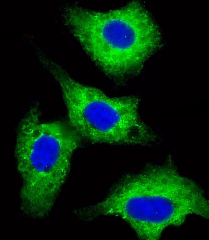
Immunofluorescent analysis of 4% paraformaldehyde-fixed, 0. 1% Triton X-100 permeabilized HepG2 (human liver hepatocellular carcinoma cell line) cells labeling Pdx1 with antibody at 1/25 dilution, followed by Dylight 488-conjugated goat anti-rabbit IgG (NK179883) secondary antibody at 1/200 dilution (green). Immunofluorescence image showing cytoplasm staining on HepG2 cell line. The nuclear counte| Product Name | DVL1 Antibody |
|---|---|
| Antibody Type | Primary Antibodies |
| Antigen Alias | Segment polarity protein dishevelled homolog DVL-1, Dishevelled-1, DSH homolog 1, DVL1 |
| Product description | DVL1, the human homolog of the Drosophila dishevelled gene (dsh) encodes a cytoplasmic phosphoprotein that regulates cell proliferation, acting as a transducer molecule for developmental processes, including segmentation and neuroblast specification. DVL1 is a candidate gene for neuroblastomatous transformation. The Schwartz-Jampel syndrome and Charcot-Marie-Tooth disease type 2A have been mapped to the same region as DVL1. The phenotypes of these diseases may be consistent with defects which might be expected from aberrant expression of a DVL gene during development.1) Metcalfe, C., et al. J. Cell. Sci. 123 (PT 9), 1588-1599 (2010) : |
| Immunogen | This DVL1 antibody is generated from rabbits immunized with a KLH conjugated synthetic peptide between 442-470 amino acids from the Central region of human DVL1. |
| Clonality | Polyclonal |
|---|---|
| Isotype | Ig |
| Host Species | Rabbit |
| Tested Applications | IFIHC-PWB |
| For IF starting dilution is: 1:25 For IHC-P starting dilution is: 1:25 For WB starting dilution is: 1:1000: |
|
| Species Reactivity | HumanMouse |
| Concentration | 1mg/ml |
| Purification | Affinity purified |
| Gene Symbol | DVL1 |
|---|---|
| Alternative Names | Segment polarity protein dishevelled homolog DVL-1 Dishevelled-1 DSH homolog 1 DVL1 |
| Molecular Weight(MW) | 75 kDa |
| Sequence Similarities | Predicted species reactivity based on immunogen sequence: Rat |
Application
Western blot analysis in MDA-MB435 cell line lysates (35ug/lane).
Application
Western Blot at 1:2000 dilution Lane 1: K562 whole cell lysates Lane 2: MDA-MB-453 whole cell lysates Lane 3: PC-3 whole cell lysates Lane 4: human kidney lysates Lysates/proteins at 20 ug per lane.
Application
Western Blot at 1:1000 dilution + PC-3 whole cell lysates Lysates/proteins at 20 ug per lane.
Application
Western Blot at 1:2000 dilution Lane 1: K562 whole cell lysate Lane 2: MDA-MB-453 whole cell lysate Lane 3: PC-3 whole cell lysate Lane 4: human kidney lysate Lane 5: NIH/3T3 whole cell lysate Lysates/proteins at 20 ug per lane.
Application
Immunofluorescent analysis of 4% paraformaldehyde-fixed, 0. 1% Triton X-100 permeabilized HepG2 (human liver hepatocellular carcinoma cell line) cells labeling Pdx1 with antibody at 1/25 dilution, followed by Dylight 488-conjugated goat anti-rabbit IgG (NK179883) secondary antibody at 1/200 dilution (green). Immunofluorescence image showing cytoplasm staining on HepG2 cell line. Cytoplasmic actin
Application
Antibody staining DVL1 in human skeletal muscle sections by Immunohistochemistry (IHC-P - paraformaldehyde-fixed, paraffin-embedded sections).
Application
Immunofluorescent analysis of 4% paraformaldehyde-fixed, 0. 1% Triton X-100 permeabilized HepG2 (human liver hepatocellular carcinoma cell line) cells labeling Pdx1 with antibody at 1/25 dilution, followed by Dylight 488-conjugated goat anti-rabbit IgG (NK179883) secondary antibody at 1/200 dilution (green). Immunofluorescence image showing cytoplasm staining on HepG2 cell line. The nuclear counte| Application Notes | For IF starting dilution is: 1:25 For IHC-P starting dilution is: 1:25 For WB starting dilution is: 1:1000: |
|---|
| Form | Liquid |
|---|---|
| Storage Instructions | Store at 4˚C for three months and -20˚C, stable for up to one year. As with all antibodies care should be taken to avoid repeated freeze thaw cycles. Antibodies should not be exposed to prolonged high temperatures. |
| Storage Buffer | Supplied in PBS with 0.09% (W/V) sodium azide. |


