Application
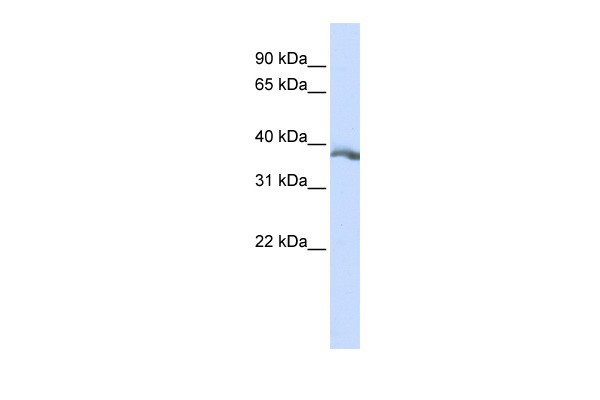
Antibody used in WB on Human HepG2 at 0.2-1 ug/ml.| Product Name | EDA Antibody |
|---|---|
| Antibody Type | Primary Antibodies |
| Antigen Alias | EDA, ED1, ED1-A1, ED1-A2, EDA1, EDA2, HED, XHED, XLHED, HED1, ODT1, ECTD1, EDA-A1, EDA-A2, STHAGX1 |
| Product description | EDA is a type II membrane protein that can be cleaved by furin to produce a secreted form. It belongs to the tumor necrosis factor family, acts as a homotrimer and may be involved in cell-cell signaling during the development of ectodermal organs. Defects in this gene are a cause of ectodermal dysplasia, anhidrotic, which is also known as X-linked hypohidrotic ectodermal dysplasia. Several transcript variants encoding many different isoforms have been found for this gene. The protein encoded by this gene is a type II membrane protein that can be cleaved by furin to produce a secreted form. The encoded protein, which belongs to the tumor necrosis factor family, acts as a homotrimer and may be involved in cell-cell signaling during the development of ectodermal organs. Defects in this gene are a cause of ectodermal dysplasia, anhidrotic, which is also known as X-linked hypohidrotic ectodermal dysplasia. Several transcript variants encoding many different isoforms have been found for this gene.1) Zhao, J., (2008) Br. J. Dermatol. 158 (3), 614-617. |
| Immunogen | Antibody produced in rabbits immunized with a synthetic peptide corresponding a region of human EDA. |
| Clonality | Polyclonal |
|---|---|
| Host Species | Rabbit |
| Tested Applications | ELISAWB |
| EDA antibody can be used for detection of EDA by ELISA at 1:312500. EDA antibody can be used for detection of EDA by western blot at 1 μg/mL, and HRP conjugated secondary antibody should be diluted 1:50,000 - 100,000.: |
|
| Species Reactivity | HumanMouse |
| Concentration | 1mg/ml |
| Purification | Affinity purified |
| Gene Symbol | EDA |
|---|---|
| Alternative Names | EDA ED1 ED1-A1 ED1-A2 EDA1 EDA2 HED XHED XLHED HED1 ODT1 ECTD1 EDA-A1 EDA-A2 STHAGX1 |
| Molecular Weight(MW) | 41 kDa |
| Entrez Gene | 1896 |
|---|---|
| Protein Accession | NP_001005609 |
Application
Antibody used in WB on Human HepG2 at 0.2-1 ug/ml.| Positive Control | 1) Cat. No. 1211 - HepG2 Cell Lysate |
|---|---|
| Application Notes | EDA antibody can be used for detection of EDA by ELISA at 1:312500. EDA antibody can be used for detection of EDA by western blot at 1 μg/mL, and HRP conjugated secondary antibody should be diluted 1:50,000 - 100,000.: |
| Form | Liquid |
|---|---|
| Storage Instructions | For short periods of storage (days) store at 4˚C. For longer periods of storage, store EDA antibody at -20˚C. As with any antibody avoid repeat freeze-thaw cycles. |
| Storage Buffer | Antibody is lyophilized in PBS buffer with 2% sucrose. Add 50 μL of distilled water. Final antibody concentration is 1 mg/mL. |


