Application
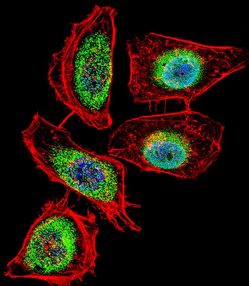
Fluorescent confocal image of Hela cell stained with ENOA Antibody (N-term).Hela cells were fixed with 4% PFA (20 min), permeabilized with Triton X-100 (0.1%, 10 min), then incubated with ENOA primary antibody (1:25). For secondary antibody, Alexa Fluor 488 conjugated donkey anti-rabbit antibody (green) was used (1:400).Cytoplasmic actin was counterstained with Alexa Fluor 555 (red) conjugated PhaApplication
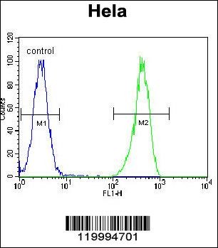
Flow cytometric analysis of Hela cells (right histogram) compared to a negative control cell (left histogram).FITC-conjugated goat-anti-rabbit secondary antibodies were used for the analysis.Application
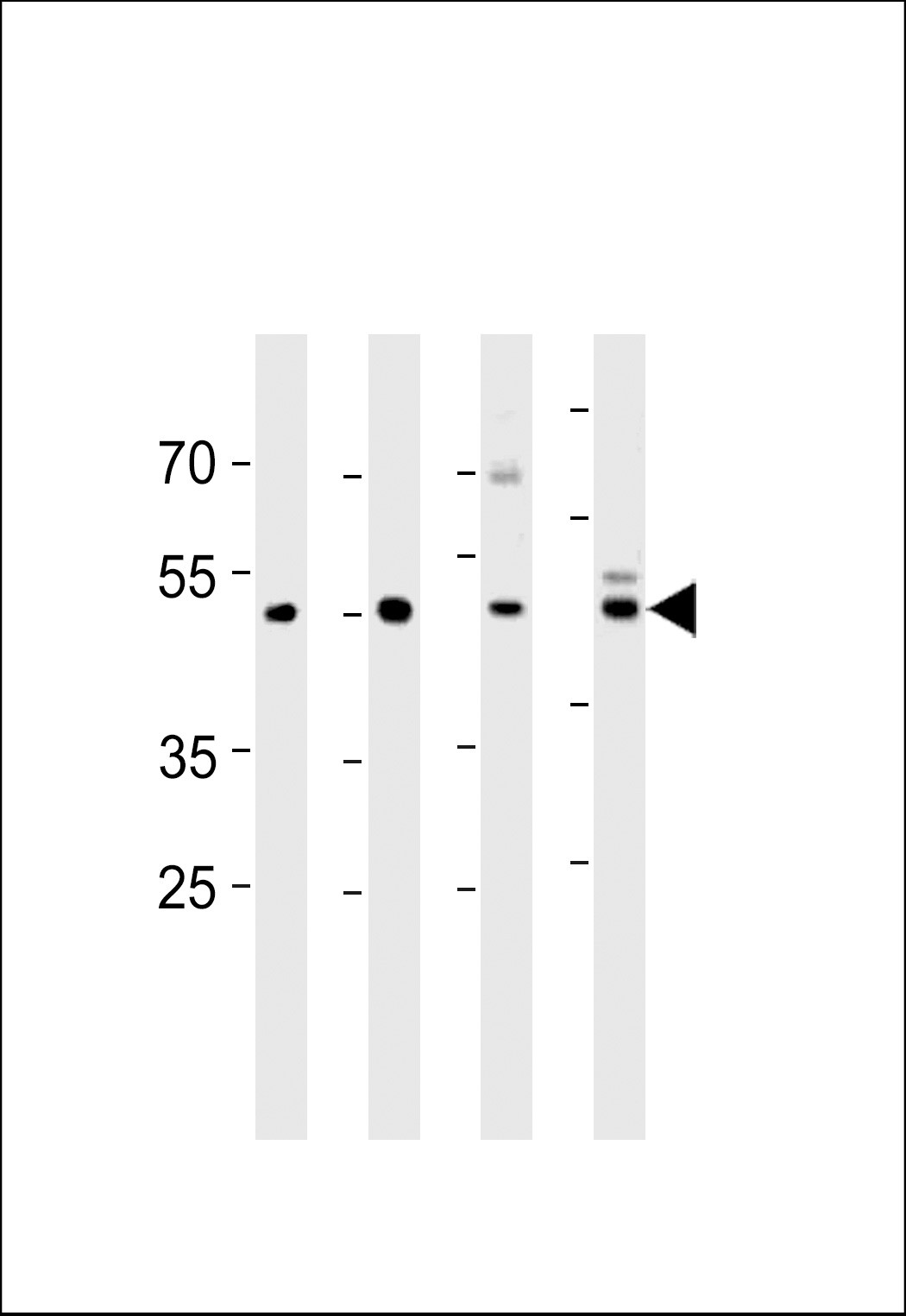
Western blot analysis in U251,mouse C2C12,mouse NIH/3T3 cell line and mouse brain tissue lysates (35ug/lane).| Product Name | ENOA Antibody |
|---|---|
| Antibody Type | Primary Antibodies |
| Antigen Alias | Alpha-enolase, 2-phospho-D-glycerate hydro-lyase, C-myc promoter-binding protein, Enolase 1, MBP-1, MPB-1, Non-neural enolase, NNE, Phosphopyruvate hydratase, Plasminogen-binding protein, ENO1, ENO1L1, MBPB1, MPB1 |
| Product description | ENO1 is one of three enolase isoenzymes found in mammals; the protein alpha-enolase, a homodimeric soluble enzyme, and is also a shorter monomeric structural lens protein, tau-crystallin. The two proteins are made from the same message. The full length protein, the isoenzyme, is found in the cytoplasm. The shorter protein is produced from an alternative translation start, is localized to the nucleus, and has been found to bind to an element in the c-myc promoter.1) Cappello,P., Int. J. Cancer 125 (3), 639-648 (2009) |
| Immunogen | This ENOA antibody is generated from rabbits immunized with a KLH conjugated synthetic peptide between 33-60 amino acids from the N-terminal region of human ENOA. |
| Clonality | Polyclonal |
|---|---|
| Isotype | Ig |
| Host Species | Rabbit |
| Tested Applications | FACSIFWB |
| For WB starting dilution is: 1:1000 For FACS starting dilution is: 1:10~50 For IF starting dilution is: 1:10~50: |
|
| Species Reactivity | HumanMouse |
| Concentration | 1mg/ml |
| Gene Symbol | ENO1 |
|---|---|
| Alternative Names | Alpha-enolase 2-phospho-D-glycerate hydro-lyase C-myc promoter-binding protein Enolase 1 MBP-1 MPB-1 Non-neural enolase NNE Phosphopyruvate hydratase Plasminogen-binding protein ENO1 ENO1L1 MBPB1 MPB1 |
| Molecular Weight(MW) | 47 kDa |
| Sequence Similarities | Predicted species reactivity based on immunogen sequence: Bovine |
Application
Fluorescent confocal image of Hela cell stained with ENOA Antibody (N-term).Hela cells were fixed with 4% PFA (20 min), permeabilized with Triton X-100 (0.1%, 10 min), then incubated with ENOA primary antibody (1:25). For secondary antibody, Alexa Fluor 488 conjugated donkey anti-rabbit antibody (green) was used (1:400).Cytoplasmic actin was counterstained with Alexa Fluor 555 (red) conjugated Pha
Application
Flow cytometric analysis of Hela cells (right histogram) compared to a negative control cell (left histogram).FITC-conjugated goat-anti-rabbit secondary antibodies were used for the analysis.
Application
Western blot analysis in U251,mouse C2C12,mouse NIH/3T3 cell line and mouse brain tissue lysates (35ug/lane).| Application Notes | For WB starting dilution is: 1:1000 For FACS starting dilution is: 1:10~50 For IF starting dilution is: 1:10~50: |
|---|
| Form | Liquid |
|---|---|
| Storage Instructions | Store at 4˚C for three months and -20˚C, stable for up to one year. As with all antibodies care should be taken to avoid repeated freeze thaw cycles. Antibodies should not be exposed to prolonged high temperatures. |
| Storage Buffer | Supplied in PBS with 0.09% (W/V) sodium azide. |


