Application
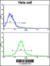
Flow cytometric analysis of hela cells using ERAS Antibody (N-term)(bottom histogram) compared to a negative control cell (top histogram). FITC-conjugated goat-anti-rabbit secondary antibodies were used for the analysis.Application
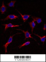
Immunofluorescence analysis of anti-ERAS Antibody in HeLa cells. 0.025 mg/ml primary antibody was followed by Alexa-Fluor-546-conjugated donkey anti-rabbit lgG (H+L). Alexa-Fluor-546 emits orange fluorescence. Blue counterstaining is DAPI.Application
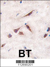
Formalin-fixed and paraffin-embedded human brain tissue reacted with ERAS antibody (A28), which was peroxidase-conjugated to the secondary antibody, followed by DAB staining.Application
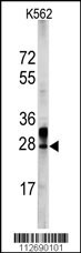
Western blot analysis of ERAS Antibody in K562 cell line lysates (35ug/lane)| Product Name | ERAS Antibody |
|---|---|
| Antibody Type | Primary Antibodies |
| Antigen Alias | GTPase ERas, E-Ras, Embryonic stem cell-expressed Ras, ERAS, HRAS2, HRASP |
| Product description | Ras proteins bind GDP/GTP and possess intrinsic GTPase activity. Point mutations of several amino acids of human RAS, including gly12, ala59, or glu63, render the protein constitutively active. Embryonic stem cell-expressed Ras (ERAS) has serine, alanine, and asparagine at the positions corresponding to gly12, ala59, and glu63 of human RAS, suggesting that it is constitutively active.The PI3K (phosphoinositide 3-kinase) pathway is important for proliferation, survival and maintenance of pluripotency in ES cells. The PI3K pathway is activated by growth factors and cytokines including insulin and leukaemia inhibitory factor. In addition to these exogenous factors, the PI3K pathway is endogenously activated by the constitutively active Ras family protein ERas (ES cell-expressed Ras). ERas null ES cells maintained pluripotency but show significantly reduced growth and tumorigenicity, which can be rescued by expression of ERas cDNA or by activated phosphatidylinositol 3-hydroxykinase. The transforming oncogene ERAS appears to be important in the tumor-like growth properties of ES cells.1) Kameda,T., Stem Cells 23 (10), 1535-1540 (2005) |
| Immunogen | This ERAS antibody is generated from rabbits immunized with a KLH conjugated synthetic peptide between 13-42 amino acids from the N-terminal region of human ERAS. |
| Clonality | Polyclonal |
|---|---|
| Isotype | Ig |
| Host Species | Rabbit |
| Tested Applications | FACSIFIHC-PWB |
| For WB starting dilution is: 1:1000 For IHC-P starting dilution is: 1:50 For IF starting dilution is: 1:10~50 For FACS starting dilution is: 1:10~50: |
|
| Species Reactivity | Human |
| Concentration | 1mg/ml |
| Gene Symbol | ERAS |
|---|---|
| Alternative Names | GTPase ERas E-Ras Embryonic stem cell-expressed Ras ERAS HRAS2 HRASP |
| Molecular Weight(MW) | 25 kDa |
Application
Flow cytometric analysis of hela cells using ERAS Antibody (N-term)(bottom histogram) compared to a negative control cell (top histogram). FITC-conjugated goat-anti-rabbit secondary antibodies were used for the analysis.
Application
Immunofluorescence analysis of anti-ERAS Antibody in HeLa cells. 0.025 mg/ml primary antibody was followed by Alexa-Fluor-546-conjugated donkey anti-rabbit lgG (H+L). Alexa-Fluor-546 emits orange fluorescence. Blue counterstaining is DAPI.
Application
Formalin-fixed and paraffin-embedded human brain tissue reacted with ERAS antibody (A28), which was peroxidase-conjugated to the secondary antibody, followed by DAB staining.
Application
Western blot analysis of ERAS Antibody in K562 cell line lysates (35ug/lane)| Application Notes | For WB starting dilution is: 1:1000 For IHC-P starting dilution is: 1:50 For IF starting dilution is: 1:10~50 For FACS starting dilution is: 1:10~50: |
|---|
| Form | Liquid |
|---|---|
| Storage Instructions | Store at 4˚C for three months and -20˚C, stable for up to one year. As with all antibodies care should be taken to avoid repeated freeze thaw cycles. Antibodies should not be exposed to prolonged high temperatures. |
| Storage Buffer | Supplied in PBS with 0.09% (W/V) sodium azide. |


