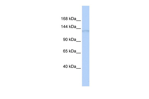
Application
Antibody used in WB on Human Brain at 0.2-1 ug/ml.| Product Name | ERCC5 Antibody |
|---|---|
| Antibody Type | Primary Antibodies |
| Antigen Alias | ERCC5, COFS3, ERCM2, UVDR, XPG, XPGC |
| Product description | Excision repair cross-complementing rodent repair deficiency, complementation group 5 (xeroderma pigmentosum, complementation group G) is involved in excision repair of UV-induced DNA damage. Mutations cause Cockayne syndrome, which is characterized by severe growth defects, mental retardation, and cachexia. Excision repair cross-complementing rodent repair deficiency, complementation group 5 (xeroderma pigmentosum, complementation group G) is involved in excision repair of UV-induced DNA damage. Mutations cause Cockayne syndrome, which is characterized by severe growth defects, mental retardation, and cachexia. Multiple alternatively spliced transcript variants encoding distinct isoforms have been described, but the biological validity of all variants has not been determined. Publication Note: This RefSeq record includes a subset of the publications that are available for this gene. Please see the Entrez Gene record to access additional publications.1) Sasaki, M., (2008) Neoplasia 10 (3), 255-265. |
| Immunogen | Antibody produced in rabbits immunized with a synthetic peptide corresponding a region of human ERCC5. |
| Clonality | Polyclonal |
|---|---|
| Host Species | Rabbit |
| Tested Applications | ELISAWB |
| ERCC5 antibody can be used for detection of ERCC5 by ELISA at 1:62500. ERCC5 antibody can be used for detection of ERCC5 by western blot at 1 μg/mL, and HRP conjugated secondary antibody should be diluted 1:50,000 - 100,000.: |
|
| Species Reactivity | HumanMouseRat |
| Concentration | 1mg/ml |
| Purification | Affinity purified |
| Gene Symbol | ERCC5 |
|---|---|
| Alternative Names | ERCC5 COFS3 ERCM2 UVDR XPG XPGC |
| Molecular Weight(MW) | 133 kDa |
Application
Antibody used in WB on Human Brain at 0.2-1 ug/ml.| Positive Control | 1) Cat. No. XBL-10123 - Fetal Brain Tissue Lysate |
|---|---|
| Application Notes | ERCC5 antibody can be used for detection of ERCC5 by ELISA at 1:62500. ERCC5 antibody can be used for detection of ERCC5 by western blot at 1 μg/mL, and HRP conjugated secondary antibody should be diluted 1:50,000 - 100,000.: |
| Form | Liquid |
|---|---|
| Storage Instructions | For short periods of storage (days) store at 4˚C. For longer periods of storage, store ERCC5 antibody at -20˚C. As with any antibody avoid repeat freeze-thaw cycles. |
| Storage Buffer | Antibody is lyophilized in PBS buffer with 2% sucrose. Add 50 μL of distilled water. Final antibody concentration is 1 mg/mL. |


