Application
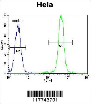
Flow cytometric analysis of Hela cells (right histogram) compared to a negative control cell (left histogram).FITC-conjugated goat-anti-rabbit secondary antibodies were used for the analysis.Application
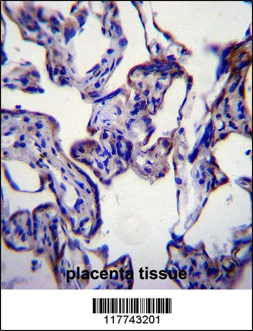
FASN Antibody immunohistochemistry analysis in formalin fixed and paraffin embedded human placenta tissue followed by peroxidase conjugation of the secondary antibody and DAB staining.Application
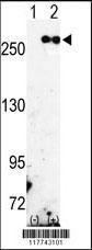
Western blot analysis of FASN using rabbit polyclonal FASN Antibody using 293 cell lysates (2 ug/lane) either nontransfected (Lane 1) or transiently transfected with the FASN gene (Lane 2).Application
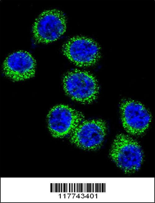
Confocal immunofluorescent analysis of FASN Antibody with Hela cell followed by Alexa Fluor 488-conjugated goat anti-rabbit lgG (green).DAPI was used to stain the cell nuclear (blue).Application
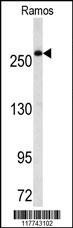
Western blot analysis of FASN Antibody in Ramos cell line lysates (35ug/lane)Application
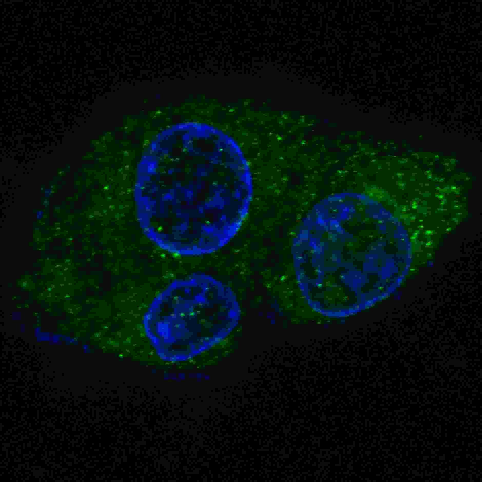
Fluorescent confocal image of HepG2 cells stained with FASN antibody. HepG2 cells were fixed with 4% PFA (20 min), permeabilized with Triton X-100 (0.2%, 30 min). Cells were then incubated with FASN primary antibody (1:200, 2 h at room temperature). For secondary antibody, Alexa Fluor 488 conjugated donkey anti-rabbit antibody (green) was used (1:1000, 1h). Nuclei were counterstained with HoechstApplication
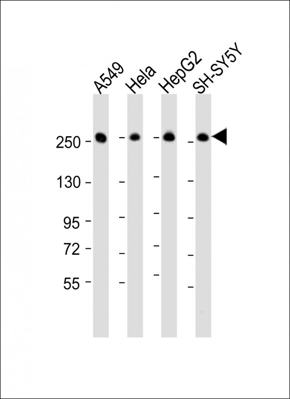
Western Blot at 1:16000 dilution Lane 1: A549 whole cell lysate Lane 2: Hela whole cell lysate Lane 3: HepG2 whole cell lysate Lane 4: SH-SY5Y whole cell lysate Lysates/proteins at 20 ug per lane.| Product Name | FASN Antibody |
|---|---|
| Antibody Type | Primary Antibodies |
| Product description | FASN is a multifunctional protein. Its main function is to catalyze the synthesis of palmitate from acetyl-CoA and malonyl-CoA, in the presence of NADPH, into long-chain saturated fatty acids. In some cancer cell lines, this protein has been found to be fused with estrogen receptor-alpha (ER-alpha), in which the N-terminus of FAS is fused in-frame with the C-terminus of ER-alpha.1) References for protein: |
| Immunogen | This FASN antibody is generated from rabbits immunized with a KLH conjugated synthetic peptide between 942-973 amino acids from the Central region of human FASN. |
| Clonality | Polyclonal |
|---|---|
| Isotype | Ig |
| Host Species | Rabbit |
| Tested Applications | FACSIFIHC-PWB |
| For WB starting dilution is: 1:16000 For IF starting dilution is: 1:200 For IHC-P starting dilution is: 1:10~50 For FACS starting dilution is: 1:10~50 | |
| Species Reactivity | Human |
| Concentration | 1mg/ml |
| Purification | Unpurified |
| Gene Symbol | FASN |
|---|---|
| Alternative Names | Fatty acid synthase [Acyl-carrier-protein] S-acetyltransferase [Acyl-carrier-protein] S-malonyltransferase 3-oxoacyl-[acyl-carrier-protein] synthase 3-oxoacyl-[acyl-carrier-protein] reductase 3-hydroxyacyl-[acyl-carrier-protein] dehydratase Enoyl-[ac |
| Molecular Weight(MW) | 273 kDa |
Application
Flow cytometric analysis of Hela cells (right histogram) compared to a negative control cell (left histogram).FITC-conjugated goat-anti-rabbit secondary antibodies were used for the analysis.
Application
FASN Antibody immunohistochemistry analysis in formalin fixed and paraffin embedded human placenta tissue followed by peroxidase conjugation of the secondary antibody and DAB staining.
Application
Western blot analysis of FASN using rabbit polyclonal FASN Antibody using 293 cell lysates (2 ug/lane) either nontransfected (Lane 1) or transiently transfected with the FASN gene (Lane 2).
Application
Confocal immunofluorescent analysis of FASN Antibody with Hela cell followed by Alexa Fluor 488-conjugated goat anti-rabbit lgG (green).DAPI was used to stain the cell nuclear (blue).
Application
Western blot analysis of FASN Antibody in Ramos cell line lysates (35ug/lane)
Application
Fluorescent confocal image of HepG2 cells stained with FASN antibody. HepG2 cells were fixed with 4% PFA (20 min), permeabilized with Triton X-100 (0.2%, 30 min). Cells were then incubated with FASN primary antibody (1:200, 2 h at room temperature). For secondary antibody, Alexa Fluor 488 conjugated donkey anti-rabbit antibody (green) was used (1:1000, 1h). Nuclei were counterstained with Hoechst
Application
Western Blot at 1:16000 dilution Lane 1: A549 whole cell lysate Lane 2: Hela whole cell lysate Lane 3: HepG2 whole cell lysate Lane 4: SH-SY5Y whole cell lysate Lysates/proteins at 20 ug per lane.| Application Notes | For WB starting dilution is: 1:16000 For IF starting dilution is: 1:200 For IHC-P starting dilution is: 1:10~50 For FACS starting dilution is: 1:10~50 |
|---|
| Form | Liquid |
|---|---|
| Storage Instructions | Store at 4˚C for three months and -20˚C, stable for up to one year. As with all antibodies care should be taken to avoid repeated freeze thaw cycles. Antibodies should not be exposed to prolonged high temperatures. |
| Storage Buffer | Supplied in PBS with 0.09% (W/V) sodium azide. |


 Data sheet for OM278570