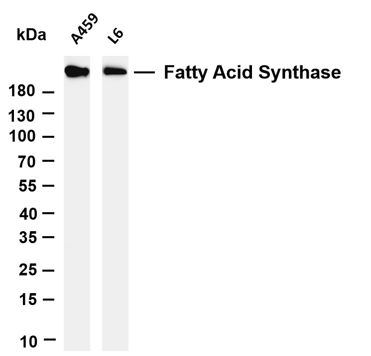
WB
Various whole cell lysates were separated by 4-20% SDS-PAGE, and the membrane was blotted with anti-Fatty Acid Synthase antibody. The HRP-conjugated Goat anti-Rabbit IgG(H + L) antibody was used to detect the antibody. Lane 1: A549, Lane 2: L6.IHC
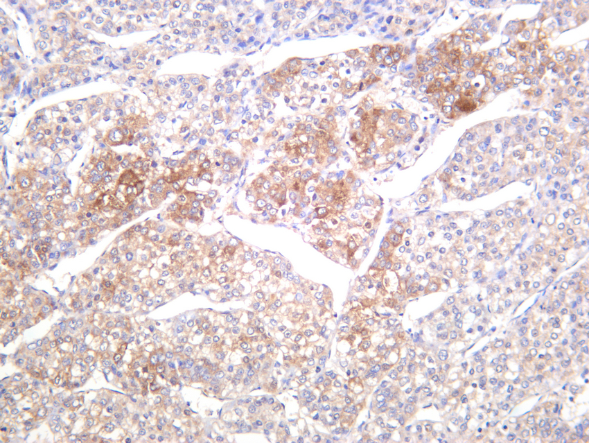
Human hepatocellular carcinoma was stained with anti-Fatty Acid Synthase rabbit antibody.IHC
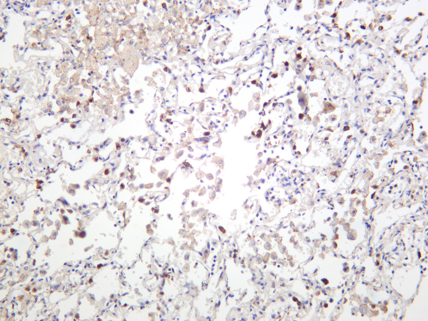
Human lung was stained with anti-Fatty Acid Synthase rabbit antibody.IHC
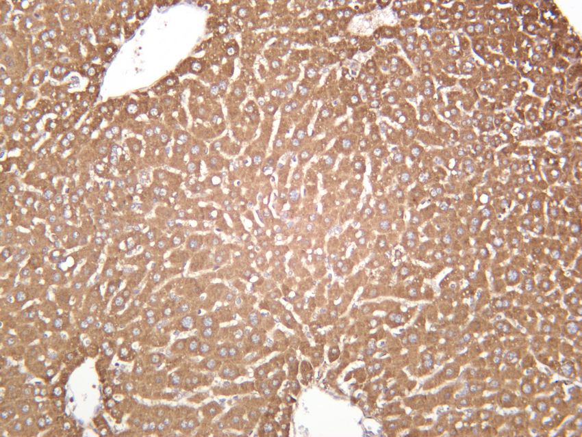
Mouse liver was stained with anti-Fatty Acid Synthase rabbit antibody.ICC/IF
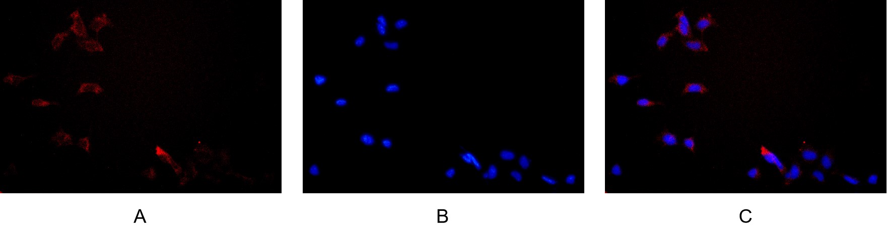
Immunofluorescence analysis of HEK293. Picture A: Fatty Acid Synthase antibody (red). Picture B: DAPI (blue). Picture C: Merge of A+B.| Product Name | Fatty Acid Synthase Rabbit mAb |
|---|---|
| Antibody Type | Primary Antibodies |
| Clonality | monoclonal |
|---|---|
| Isotype | IgG |
| Host Species | Rabbit |
| Tested Applications | ICC/IFIHCWB |
| WB:1:2000-1:10000 IHC:1:500-1:2000 ICC/IF:1:200-1:1000 |
|
| Species Reactivity | HumanMouseRat |
| Concentration | 1mg/ml |
| Purification | Protein A |
| Gene Symbol | FASN |
|---|---|
| Gene Synonyms | FAS OA-519 SDR27X1 |
| Gene Full Name | fatty acid synthase |
| Gene Summary | The enzyme encoded by this gene is a multifunctional protein. Its main function is to catalyze the synthesis of palmitate from acetyl-CoA and malonyl-CoA, in the presence of NADPH, into long-chain saturated fatty acids. In some cancer cell lines, this protein has been found to be fused with estrogen receptor-alpha (ER-alpha), in which the N-terminus of FAS is fused in-frame with the C-terminus of ER-alpha. [provided by RefSeq, Jul 2008] |
| Molecular Weight(MW) | 273kDa |
| Cellular Localization | Cytoplasm. |
WB
Various whole cell lysates were separated by 4-20% SDS-PAGE, and the membrane was blotted with anti-Fatty Acid Synthase antibody. The HRP-conjugated Goat anti-Rabbit IgG(H + L) antibody was used to detect the antibody. Lane 1: A549, Lane 2: L6.
IHC
Human hepatocellular carcinoma was stained with anti-Fatty Acid Synthase rabbit antibody.
IHC
Human lung was stained with anti-Fatty Acid Synthase rabbit antibody.
IHC
Mouse liver was stained with anti-Fatty Acid Synthase rabbit antibody.
ICC/IF
Immunofluorescence analysis of HEK293. Picture A: Fatty Acid Synthase antibody (red). Picture B: DAPI (blue). Picture C: Merge of A+B.| Application Notes | WB:1:2000-1:10000 IHC:1:500-1:2000 ICC/IF:1:200-1:1000 |
|---|
| Form | Liquid |
|---|---|
| Storage Instructions | -15°C to -25°C/1 year(Do not lower than -25°C) |
| Storage Buffer | PBS, 50% glycerol, 0.05% Proclin 300, 0.05%BSA |


 Data sheet for OM644289