Application
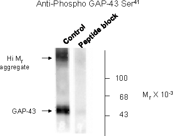
Western blot of rat cortex lysate showing phospho-specific immunolabeling of the ~50k GAP-43 protein phosphorylated at Ser41.| Product Name | Gap-43 (phospho Ser41) Antibody |
|---|---|
| Antibody Type | Primary Antibodies |
| Antigen Alias | Basp2, Axonal membrane protein GAP-43, |
| Product description | GAP-43 is thought to have an important role in development and plasticity because it is expressed at high levels in neuronal growth cones during development and during axonal regeneration. There is also evidence from knockout animals that GAP-43 serves to amplify pathfinding signals from the growth cone. GAP-43 is thought to mediate at least some of these effects via interaction with actin. Importantly, phosphorylation at Ser41 by Protein Kinase C modulates the interaction of GAP-43 with actin.1) Hulo, S., Alberi, S., Laux, T., Muller, D., and Caroni, P., "A point mutant of GAP-43 induces enhanced short-term and long-term hippocampal plasticity," Eur. J. Neurosci., 15 (2002) 1976 - 1982. |
| Immunogen | GAP-43 (Ser41) polyclonal antibody was raised against a synthetic phosphopeptide corresponding to amino acids residues surrounding the phospho-Ser41 of rat GAP-43. |
| Modification | p-Ser41 |
| Clonality | Polyclonal |
|---|---|
| Host Species | Rabbit |
| Tested Applications | WB |
| In some tissues the antibody also recognizes a higher molecular weight band that is also recognized by the Pan GAP-43 antibody and this band may be a GAP-43 aggregate or oligomer. Immunolabeling of the GAP-43 protein in Western blots of rat brain is blocked by the Ser41 phosphopeptide used as antigen but not by the corresponding non-phosphopeptide. Note: the GAP-43 protein migrates on SDS-PAGE as a 50 kDa band. The antibody is purified by sequential chromatography on phospho- and non-phosphopeptide affinity columns. Antibody dilutions and tissue load should be based on tissue type and expected phosphorylation state. Initial recommended range of dilutions: 1:500 to 1:2000. Applications include Dot blots (DB) and Western blots (WB). Immunohistochemistry (IHC) application has not yet been determined. Human, mouse and rat have 100% amino acid sequence identity with for the antigen used to raise the antibody. When internally tested under ideal conditions the working dilutions were 1:1000 for DB and WB.: |
|
| Species Reactivity | HumanMouseRat |
| Concentration | 1mg/ml |
| Purification | Affinity purified |
| Gene Symbol | Gap43 |
|---|---|
| Alternative Names | Basp2 Axonal membrane protein GAP-43 |
| Molecular Weight(MW) | 50 |
| Tissue Specificity | Gap-43 antibody is specific for GAP-43 phosphorylated at Ser41. |
Application
Western blot of rat cortex lysate showing phospho-specific immunolabeling of the ~50k GAP-43 protein phosphorylated at Ser41.| Application Notes | In some tissues the antibody also recognizes a higher molecular weight band that is also recognized by the Pan GAP-43 antibody and this band may be a GAP-43 aggregate or oligomer. Immunolabeling of the GAP-43 protein in Western blots of rat brain is blocked by the Ser41 phosphopeptide used as antigen but not by the corresponding non-phosphopeptide. Note: the GAP-43 protein migrates on SDS-PAGE as a 50 kDa band. The antibody is purified by sequential chromatography on phospho- and non-phosphopeptide affinity columns. Antibody dilutions and tissue load should be based on tissue type and expected phosphorylation state. Initial recommended range of dilutions: 1:500 to 1:2000. Applications include Dot blots (DB) and Western blots (WB). Immunohistochemistry (IHC) application has not yet been determined. Human, mouse and rat have 100% amino acid sequence identity with for the antigen used to raise the antibody. When internally tested under ideal conditions the working dilutions were 1:1000 for DB and WB.: |
|---|
| Form | Liquid |
|---|---|
| Storage Instructions | For long term storage -80˚C is recommended, but shorter term storage at -20˚C is also acceptable as aliquots may be taken without freeze/thawing due to the presence of 50% glycerol. Stock solutions are stable for a minimum of 1 year at -20˚C. |
| Storage Buffer | 100 uL in 10 mM HEPES (pH 7.5), 150 mM NaCl, 100 ug per mL BSA and 50% glycerol. |


