Application
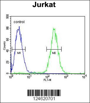
Flow cytometric analysis of Jurkat cells (right histogram) compared to a negative control cell (left histogram).FITC-conjugated goat-anti-rabbit secondary antibodies were used for the analysis.Application
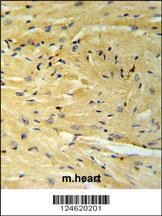
GNAT1 Antibody IHC analysis in formalin fixed and paraffin embedded moouse heart tissue followed by peroxidase conjugation of the secondary antibody and DAB staining.Application
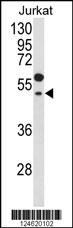
Western blot analysis of GNAT1 Antibody in Jurkat cell line lysates (35ug/lane)| Product Name | GNAT1 Antibody |
|---|---|
| Antibody Type | Primary Antibodies |
| Antigen Alias | Guanine nucleotide-binding protein G(t) subunit alpha-1, Transducin alpha-1 chain, GNAT1, GNATR |
| Product description | Transducin is a 3-subunit guanine nucleotide-binding protein (G protein) which stimulates the coupling of rhodopsin and cGMP-phoshodiesterase during visual impulses. The transducin alpha subunits in rods and cones are encoded by separate genes. This gene encodes the alpha subunit in rods. This gene is also expressed in other cells, and has been implicated in bitter taste transduction in rat taste cells.1) Luttrell, L.M. Mol. Biotechnol. 39(3):239-264(2008) |
| Immunogen | This GNAT1 antibody is generated from rabbits immunized with a KLH conjugated synthetic peptide between 290-318 amino acids from the C-terminal region of human GNAT1. |
| Clonality | Polyclonal |
|---|---|
| Isotype | Ig |
| Host Species | Rabbit |
| Tested Applications | FACSIHC-PWB |
| For WB starting dilution is: 1:1000 For IHC-P starting dilution is: 1:50~100 For FACS starting dilution is: 1:10~50: |
|
| Species Reactivity | Human |
| Concentration | 1mg/ml |
| Purification | Affinity purified |
| Gene Symbol | GNAT1 |
|---|---|
| Alternative Names | Guanine nucleotide-binding protein G(t) subunit alpha-1 Transducin alpha-1 chain GNAT1 GNATR |
| Molecular Weight(MW) | 40 kDa |
| Sequence Similarities | Predicted species reactivity based on immunogen sequence: Bovine |
Application
Flow cytometric analysis of Jurkat cells (right histogram) compared to a negative control cell (left histogram).FITC-conjugated goat-anti-rabbit secondary antibodies were used for the analysis.
Application
GNAT1 Antibody IHC analysis in formalin fixed and paraffin embedded moouse heart tissue followed by peroxidase conjugation of the secondary antibody and DAB staining.
Application
Western blot analysis of GNAT1 Antibody in Jurkat cell line lysates (35ug/lane)| Application Notes | For WB starting dilution is: 1:1000 For IHC-P starting dilution is: 1:50~100 For FACS starting dilution is: 1:10~50: |
|---|
| Form | Liquid |
|---|---|
| Storage Instructions | Store at 4˚C for three months and -20˚C, stable for up to one year. As with all antibodies care should be taken to avoid repeated freeze thaw cycles. Antibodies should not be exposed to prolonged high temperatures. |
| Storage Buffer | Supplied in PBS with 0.09% (W/V) sodium azide. |


