Application
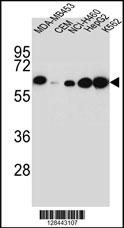
Western blot analysis in MDA-MB453,CEM,NCI-H460,HepG2,K562 cell line lysates (35ug/lane).This demonstrates the detected the HP protein (arrow).Application
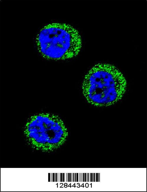
Confocal immunofluorescent analysis of HP Antibody with MDA-MB435 cell followed by Alexa Fluor 488-conjugated goat anti-rabbit lgG (green).DAPI was used to stain the cell nuclear (blue).Application
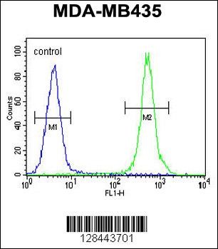
Flow cytometric analysis of MDA-MB435 cells (right histogram) compared to a negative control cell (left histogram).FITC-conjugated goat-anti-rabbit secondary antibodies were used for the analysis.Application
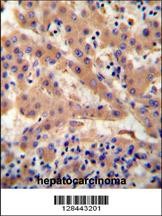
HP antibody immunohistochemistry analysis in formalin fixed and paraffin embedded human hepatocarcinoma followed by peroxidase conjugation of the secondary antibody and DAB staining.Application
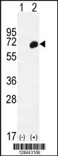
Western blot analysis of HP using rabbit polyclonal HP Antibody using 293 cell lysates (2 ug/lane) either nontransfected (Lane 1) or transiently transfected (Lane 2) with the HP gene.Application
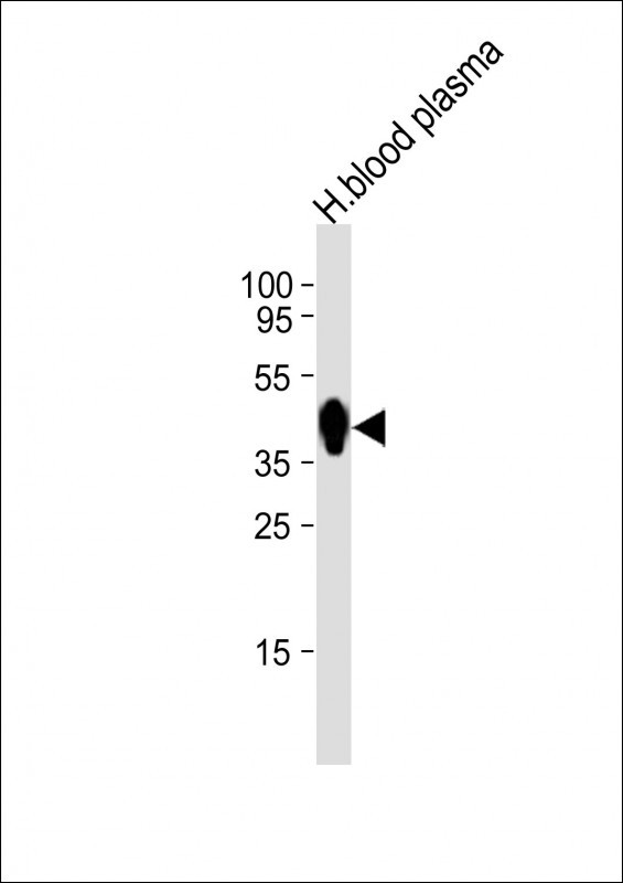
Western Blot at 1:1000 dilution + human blood plasma lysates Lysates/proteins at 20 ug per lane.| Product Name | HP Antibody |
|---|---|
| Antibody Type | Primary Antibodies |
| Antigen Alias | Haptoglobin, Zonulin, Haptoglobin alpha chain, Haptoglobin beta chain, HP |
| Product description | This gene encodes a preproprotein, which is processed to yield both alpha and beta chains, which subsequently combine as a tetramer to produce haptoglobin. Haptoglobin functions to bind free plasma hemoglobin, which allows degradative enzymes to gain access to the hemoglobin, while at the same time preventing loss of iron through the kidneys and protecting the kidneys from damage by hemoglobin. Mutations in this gene and/or its regulatory regions cause ahaptoglobinemia or hypohaptoglobinemia. This gene has also been linked to diabetic nephropathy, the incidence of coronary artery disease in type 1 diabetes, Crohn's disease, inflammatory disease behavior, primary sclerosing cholangitis, susceptibility to idiopathic Parkinson's disease, and a reduced incidence of Plasmodium falciparum malaria. A similar duplicated gene is located next to this gene on chromosome 16. Multiple transcript variants encoding different isoforms have been found for this gene.1) Bailey, S.D., et al. Diabetes Care 33(10):2250-2253(2010) |
| Immunogen | This HP antibody is generated from rabbits immunized with a KLH conjugated synthetic peptide between 296-322 amino acids from the Central region of human HP. |
| Clonality | Polyclonal |
|---|---|
| Isotype | Ig |
| Host Species | Rabbit |
| Tested Applications | FACSIFIHC-PWB |
| For WB starting dilution is: 1:1000 For IHC-P starting dilution is: 1:50~100 For FACS starting dilution is: 1:10~50 For IF starting dilution is: 1:10~50: |
|
| Species Reactivity | Human |
| Concentration | 1mg/ml |
| Purification | Affinity purified |
| Gene Symbol | HP |
|---|---|
| Alternative Names | Haptoglobin Zonulin Haptoglobin alpha chain Haptoglobin beta chain HP |
| Molecular Weight(MW) | 45 kDa |
Application
Western blot analysis in MDA-MB453,CEM,NCI-H460,HepG2,K562 cell line lysates (35ug/lane).This demonstrates the detected the HP protein (arrow).
Application
Confocal immunofluorescent analysis of HP Antibody with MDA-MB435 cell followed by Alexa Fluor 488-conjugated goat anti-rabbit lgG (green).DAPI was used to stain the cell nuclear (blue).
Application
Flow cytometric analysis of MDA-MB435 cells (right histogram) compared to a negative control cell (left histogram).FITC-conjugated goat-anti-rabbit secondary antibodies were used for the analysis.
Application
HP antibody immunohistochemistry analysis in formalin fixed and paraffin embedded human hepatocarcinoma followed by peroxidase conjugation of the secondary antibody and DAB staining.
Application
Western blot analysis of HP using rabbit polyclonal HP Antibody using 293 cell lysates (2 ug/lane) either nontransfected (Lane 1) or transiently transfected (Lane 2) with the HP gene.
Application
Western Blot at 1:1000 dilution + human blood plasma lysates Lysates/proteins at 20 ug per lane.| Application Notes | For WB starting dilution is: 1:1000 For IHC-P starting dilution is: 1:50~100 For FACS starting dilution is: 1:10~50 For IF starting dilution is: 1:10~50: |
|---|
| Form | Liquid |
|---|---|
| Storage Instructions | Store at 4˚C for three months and -20˚C, stable for up to one year. As with all antibodies care should be taken to avoid repeated freeze thaw cycles. Antibodies should not be exposed to prolonged high temperatures. |
| Storage Buffer | Supplied in PBS with 0.09% (W/V) sodium azide. |


