Application
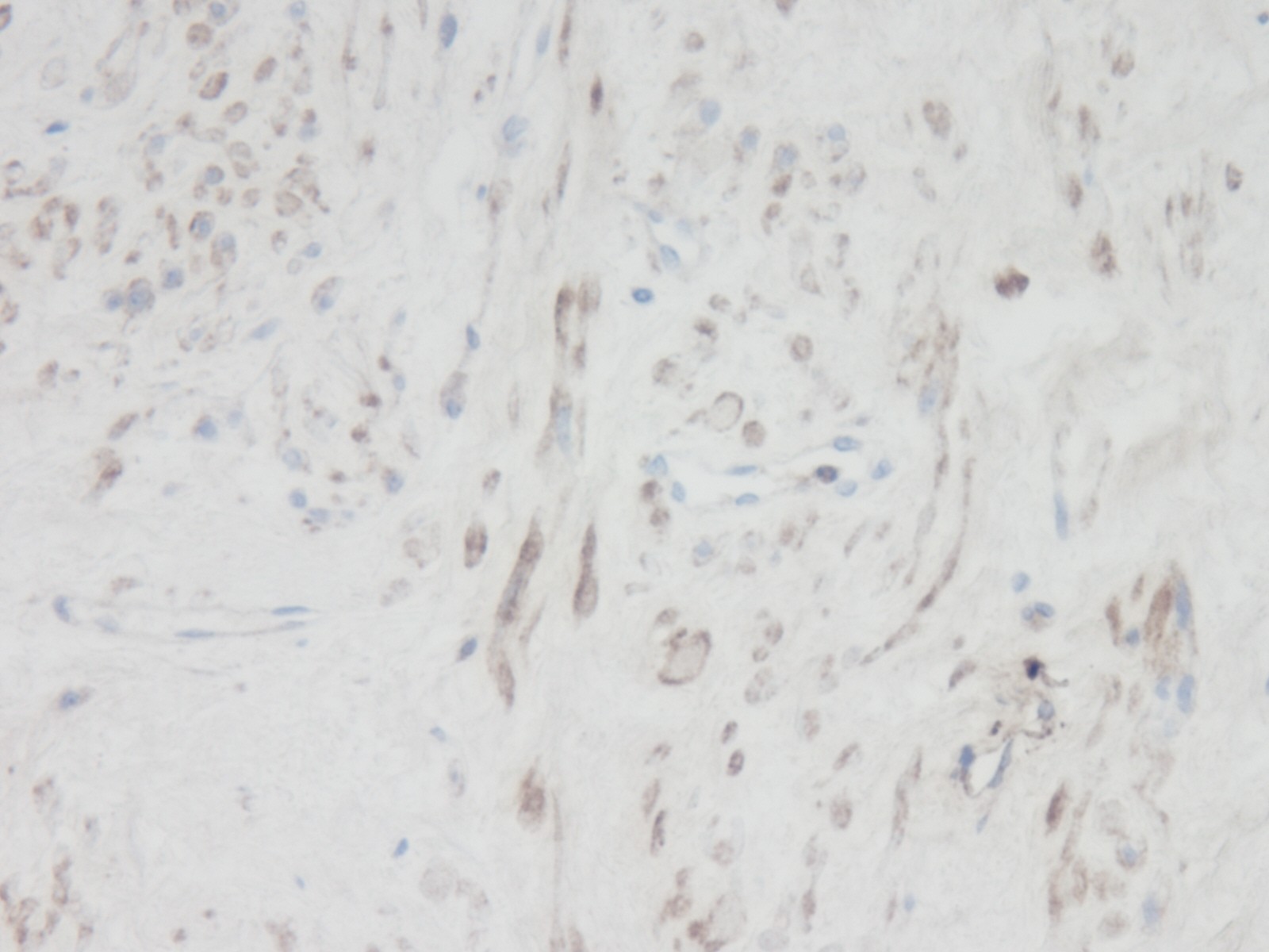
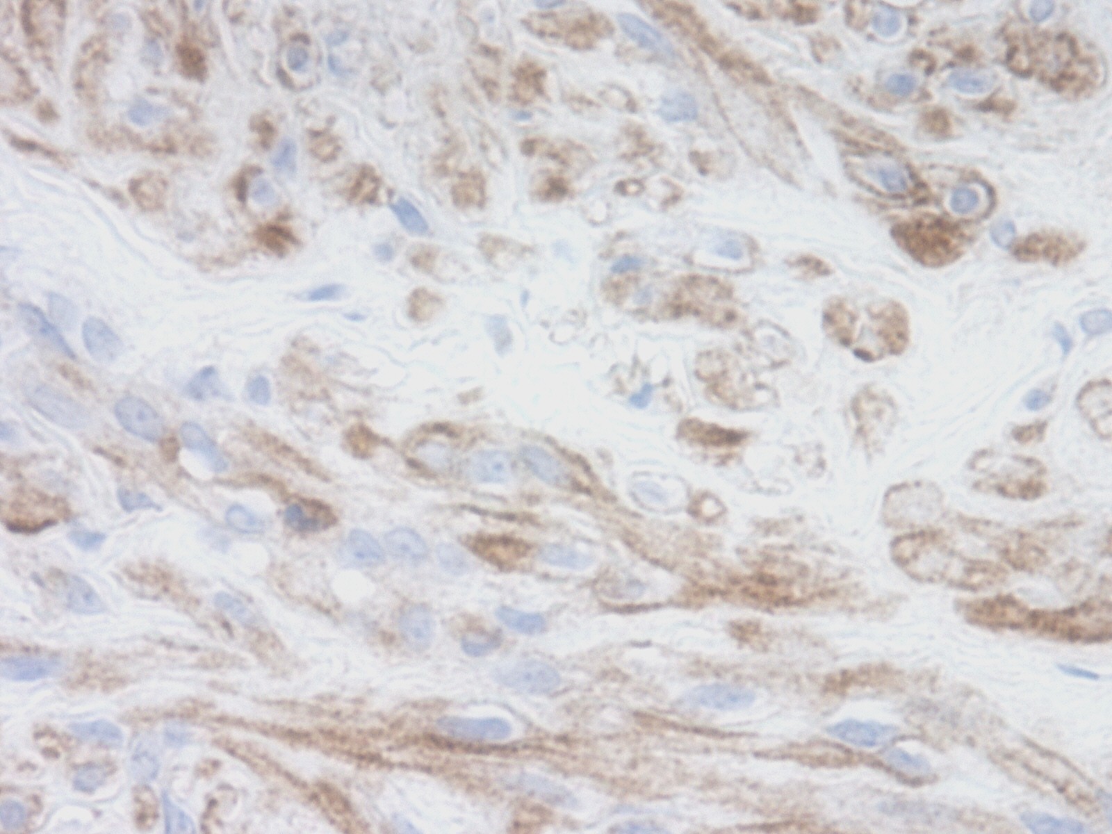
This antibody stained formalin-fixed paraffin-embedded sections of human renal tumor with parenchyma tissue. The recommended concentration is 0.125ug/ml- 0.25ug/ml with an overnight incubation at 4˚C. An HRP-labeled polymer detection system was used with a DAB chromogen. Optimal results for these conditions were achieved without antigen retrieval. Optimal concentrations and conditions may varApplication
This antibody stained formalin-fixed paraffin-embedded sections of human renal tumor with parenchyma tissue. The recommended concentration is 0.125ug/ml- 0.25ug/ml with an overnight incubation at 4˚C. An HRP-labeled polymer detection system was used with a DAB chromogen. Optimal results for these conditions were achieved without antigen retrieval. Optimal concentrations and conditions may varApplication
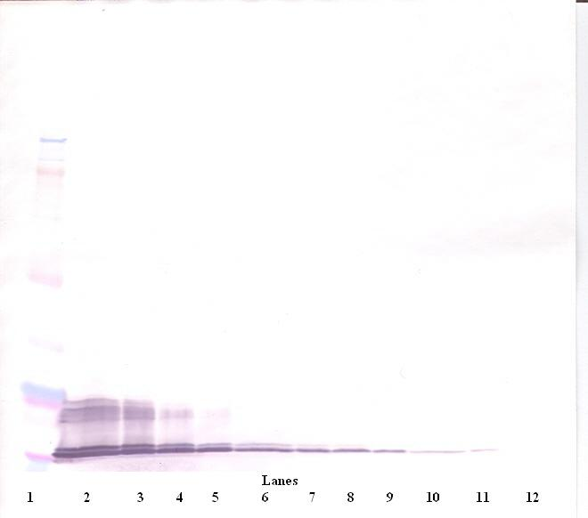
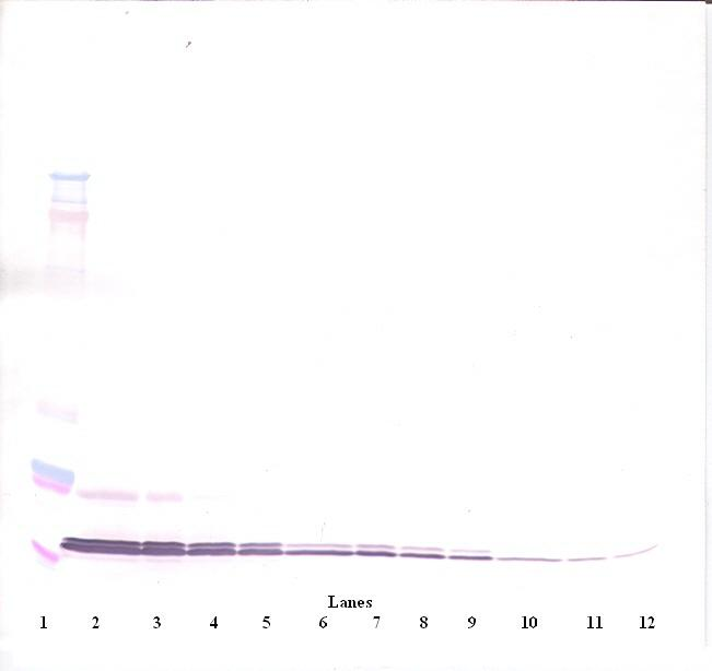
To detect hI-TAC by Western Blot analysis this antibody can be used at a concentration of 0.1-0.2 ug/ml. Used in conjunction with compatible secondary reagents the detection limit for recombinant hI-TAC is 1.5-3.0 ng/lane, under either reducing or non-reducing conditions.Application
To detect hI-TAC by Western Blot analysis this antibody can be used at a concentration of 0.1-0.2 ug/ml. Used in conjunction with compatible secondary reagents the detection limit for recombinant hI-TAC is 1.5-3.0 ng/lane, under either reducing or non-reducing conditions.Application
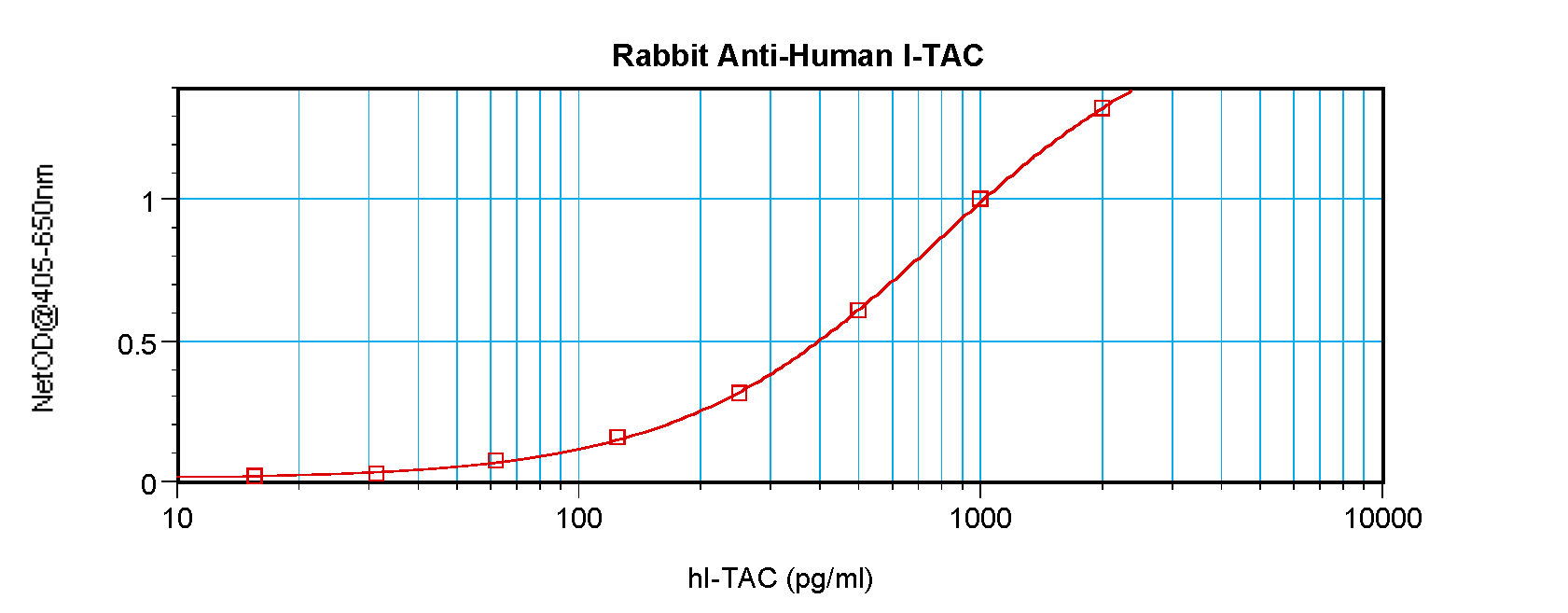
To detect hI-TAC by sandwich ELISA (using 100 ul/well antibody solution) a concentration of 0.5 - 2.0 ug/ml of this antibody is required. This antigen affinity purified antibody, in conjunction with ProSci’s Biotinylated Anti-Human I-TAC (XP-5205Bt) as a detection antibody, allows the detection of at least 0.2 - 0.4 ng/well of recombinant hI-TAC.| Product Name | I-TAC Antibody |
|---|---|
| Antibody Type | Primary Antibodies |
| Antigen Alias | IP9, H174, IP-9, b-R1, I-TAC, SCYB11, SCYB9B, ITAC, C-X-C motif chemokine 11, Beta-R1 |
| Immunogen | Produced from sera of rabbits pre-immunized with highly pure (>98%) recombinant hI-TAC (human Interferon-Inducible T Cell Alpha Chemoattractant). |
| Clonality | Polyclonal |
|---|---|
| Host Species | Rabbit |
| Tested Applications | ELISANeutWB |
| Neutralization: To yield one-half maximal inhibition [ND50] of the biological activity of hI-TAC (100 ng/mL), a concentration of 2.0 - 3.0 μg/mL of this antibody is required. ELISA: To detect hI-TAC by direct ELISA (using 100 μL/well antibody solution) a concentration of at least 0.5 μg/mL of this antibody is required. This antigen affinity purified antibody, in conjunction with compatible secondary reagents, allows the detection of 0.2 - 0.4 ng/well of recombinant hI-TAC. Sandwich: To detect hI-TAC by sandwich ELISA (using 100 μL/well antibody solution) a concentration of 0.5 - 2.0 μg/mL of this antibody is required. This antigen affinity purified antibody, in conjunction with our Biotinylated Anti-Human I-TAC (XP-5205Bt) as a detection antibody, allows the detection of at least 0.2 - 0.4 ng/well of recombinant hI-TAC. Western Blot: To detect hI-TAC by Western Blot analysis this antibody can be used at a concentration of 0.1 - 0.2 μg/mL. Used in conjunction with compatible secondary reagents the detection limit for recombinant hI-TAC is 1.5 - 3.0 ng/lane, under either reducing or non-reducing conditions.: |
|
| Species Reactivity | Human |
| Concentration | 1mg/ml |
| Purification | Affinity purified |
| Gene Symbol | CXCL11 |
|---|---|
| Alternative Names | IP9 H174 IP-9 b-R1 I-TAC SCYB11 SCYB9B ITAC C-X-C motif chemokine 11 Beta-R1 |
Application
This antibody stained formalin-fixed paraffin-embedded sections of human renal tumor with parenchyma tissue. The recommended concentration is 0.125ug/ml- 0.25ug/ml with an overnight incubation at 4˚C. An HRP-labeled polymer detection system was used with a DAB chromogen. Optimal results for these conditions were achieved without antigen retrieval. Optimal concentrations and conditions may var
Application
This antibody stained formalin-fixed paraffin-embedded sections of human renal tumor with parenchyma tissue. The recommended concentration is 0.125ug/ml- 0.25ug/ml with an overnight incubation at 4˚C. An HRP-labeled polymer detection system was used with a DAB chromogen. Optimal results for these conditions were achieved without antigen retrieval. Optimal concentrations and conditions may var
Application
To detect hI-TAC by Western Blot analysis this antibody can be used at a concentration of 0.1-0.2 ug/ml. Used in conjunction with compatible secondary reagents the detection limit for recombinant hI-TAC is 1.5-3.0 ng/lane, under either reducing or non-reducing conditions.
Application
To detect hI-TAC by Western Blot analysis this antibody can be used at a concentration of 0.1-0.2 ug/ml. Used in conjunction with compatible secondary reagents the detection limit for recombinant hI-TAC is 1.5-3.0 ng/lane, under either reducing or non-reducing conditions.
Application
To detect hI-TAC by sandwich ELISA (using 100 ul/well antibody solution) a concentration of 0.5 - 2.0 ug/ml of this antibody is required. This antigen affinity purified antibody, in conjunction with ProSci’s Biotinylated Anti-Human I-TAC (XP-5205Bt) as a detection antibody, allows the detection of at least 0.2 - 0.4 ng/well of recombinant hI-TAC.| Application Notes | Neutralization: To yield one-half maximal inhibition [ND50] of the biological activity of hI-TAC (100 ng/mL), a concentration of 2.0 - 3.0 μg/mL of this antibody is required. ELISA: To detect hI-TAC by direct ELISA (using 100 μL/well antibody solution) a concentration of at least 0.5 μg/mL of this antibody is required. This antigen affinity purified antibody, in conjunction with compatible secondary reagents, allows the detection of 0.2 - 0.4 ng/well of recombinant hI-TAC. Sandwich: To detect hI-TAC by sandwich ELISA (using 100 μL/well antibody solution) a concentration of 0.5 - 2.0 μg/mL of this antibody is required. This antigen affinity purified antibody, in conjunction with our Biotinylated Anti-Human I-TAC (XP-5205Bt) as a detection antibody, allows the detection of at least 0.2 - 0.4 ng/well of recombinant hI-TAC. Western Blot: To detect hI-TAC by Western Blot analysis this antibody can be used at a concentration of 0.1 - 0.2 μg/mL. Used in conjunction with compatible secondary reagents the detection limit for recombinant hI-TAC is 1.5 - 3.0 ng/lane, under either reducing or non-reducing conditions.: |
|---|
| Form | Liquid |
|---|---|
| Storage Instructions | I-TAC antibody is stable for at least 2 years from date of receipt at -20˚C. The reconstituted antibody is stable for at least two weeks at 2-8˚C. Frozen aliquots are stable for at least 6 months when stored at -20˚C. Avoid repeated freeze-thaw cycles. |
| Storage Buffer | phosphate buffered saline , pH 7.4, 150mM NaCl, 0.02% sodium azide and 50% glycerol. |


