
Application
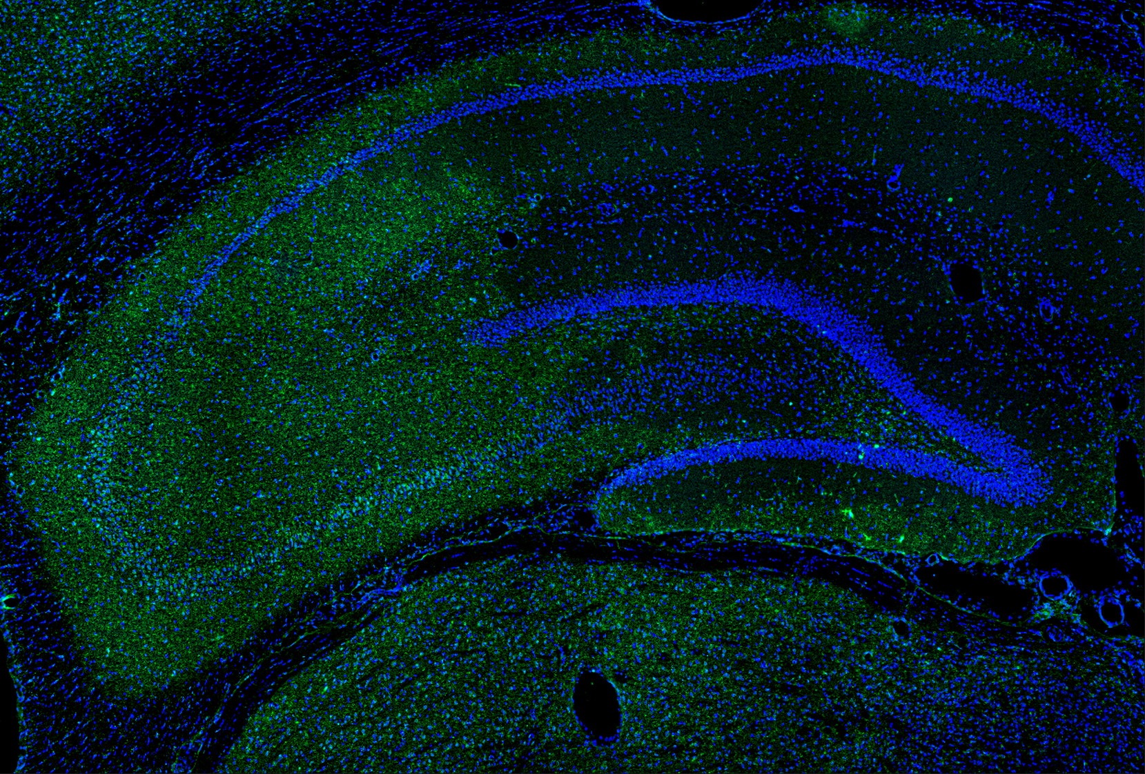
This antibody stained colchicine injected rat brain (including the ventricles and the CA3 region of the hippocampus) tissue. The primary antibody was incubated at 0.25 ug/ml overnight at 4˚C. This was followed by a peroxidase conjugated secondary antibody and then a fluorescein Tyramide Signal Amplification (TSA) reagent. Optimal concentrations and conditions may vary.Application
This antibody stained colchicine injected rat brain (including the ventricles and the CA3 region of the hippocampus) tissue. The primary antibody was incubated at 0.25 ug/ml overnight at 4˚C. This was followed by a peroxidase conjugated secondary antibody and then a fluorescein Tyramide Signal Amplification (TSA) reagent. Optimal concentrations and conditions may vary.Application
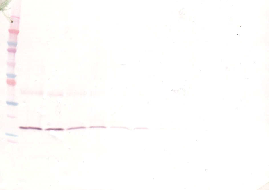
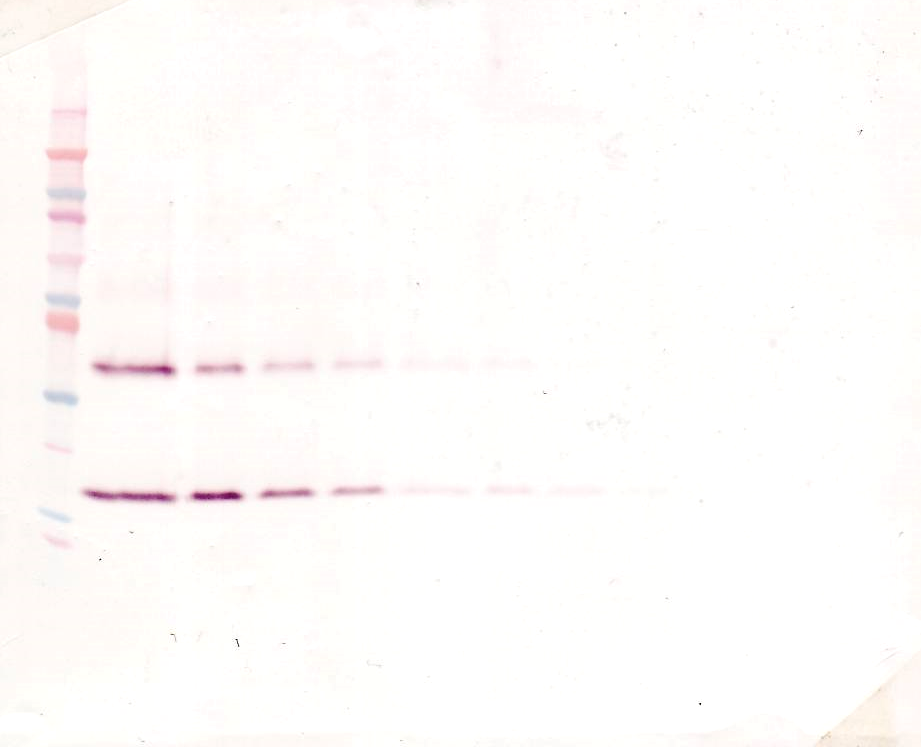
To detect Rat IL-1-beta by Western Blot analysis this antibody can be used at a concentration of 0.1-0.2 ug/ml. When used in conjunction with compatible secondary reagents, the detection limit for recombinant Rat IL-1-beta is 1.5-3.0 ng/lane, under either reducing or non-reducing conditions.Application
To detect Rat IL-1-beta by Western Blot analysis this antibody can be used at a concentration of 0.1-0.2 ug/ml. When used in conjunction with compatible secondary reagents, the detection limit for recombinant Rat IL-1-beta is 1.5-3.0 ng/lane, under either reducing or non-reducing conditions.Application
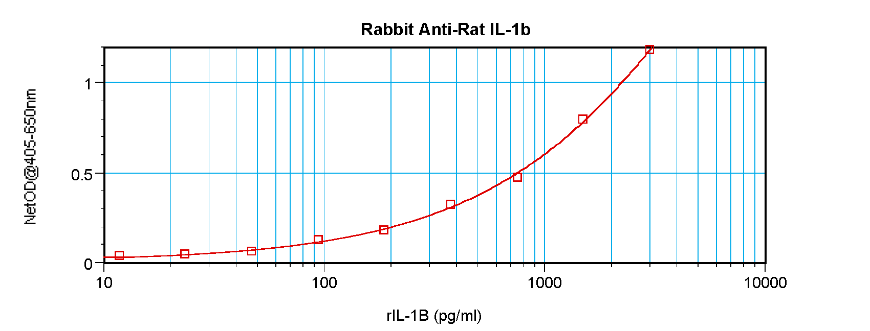
To detect Rat IL-1-beta by sandwich ELISA (using 100 ul/well antibody solution) a concentration of 0.5 - 2.0 ug/ml of this antibody is required. This antigen affinity purified antibody, in conjunction with ProSci’s Biotinylated Anti-Rat IL-1-beta (XP-5180Bt) as a detection antibody, allows the detection of at least 0.2 - 0.4 ng/well of recombinant Rat IL-1-beta.| Product Name | IL-1B Antibody |
|---|---|
| Antibody Type | Primary Antibodies |
| Antigen Alias | Interleukin-1 betaIL-1 beta |
| Immunogen | Produced from sera of rabbits pre-immunized with highly pure (>98%) recombinant rIL-1-beta (rat Interleukin-1-beta). |
| Clonality | Polyclonal |
|---|---|
| Host Species | Rabbit |
| Tested Applications | ELISANeutWB |
| Neutralization: To yield one-half maximal inhibition [ND50] of the biological activity of rIL-1-beta (0.30 ng/mL), a concentration of 0.003 - 0.005 μg/mL of this antibody is required. ELISA: To detect rIL-1-beta by direct ELISA (using 100 μL/well antibody solution) a concentration of at least 0.5 μg/mL of this antibody is required. This antigen affinity purified antibody, in conjunction with compatible secondary reagents, allows the detection of 0.2 - 0.4 ng/well of recombinant rIL-1-beta. Sandwich: To detect Rat IL-1β by sandwich ELISA (using 100 μL/well antibody solution) a concentration of 0.5 - 2.0 μg/mL of this antibody is required. This antigen affinity purified antibody, in conjunction with our Biotinylated Anti-Rat IL-1β (XP-5180Bt) as a detection antibody, allows the detection of at least 0.2 - 0.4 ng/well of recombinant Rat IL-1β. Western Blot: To detect rIL-1-beta by Western Blot analysis this antibody can be used at a concentration of 0.1 - 0.2 μg/mL. Used in conjunction with compatible secondary reagents the detection limit for recombinant rIL-1-beta is 1.5 - 3.0 ng/lane, under either reducing or non-reducing conditions.: |
|
| Species Reactivity | Rat |
| Concentration | 1mg/ml |
| Purification | Affinity purified |
| Gene Symbol | Il1b |
|---|---|
| Alternative Names | Interleukin-1 betaIL-1 beta |
Application
This antibody stained colchicine injected rat brain (including the ventricles and the CA3 region of the hippocampus) tissue. The primary antibody was incubated at 0.25 ug/ml overnight at 4˚C. This was followed by a peroxidase conjugated secondary antibody and then a fluorescein Tyramide Signal Amplification (TSA) reagent. Optimal concentrations and conditions may vary.
Application
To detect Rat IL-1-beta by Western Blot analysis this antibody can be used at a concentration of 0.1-0.2 ug/ml. When used in conjunction with compatible secondary reagents, the detection limit for recombinant Rat IL-1-beta is 1.5-3.0 ng/lane, under either reducing or non-reducing conditions.
Application
To detect Rat IL-1-beta by sandwich ELISA (using 100 ul/well antibody solution) a concentration of 0.5 - 2.0 ug/ml of this antibody is required. This antigen affinity purified antibody, in conjunction with ProSci’s Biotinylated Anti-Rat IL-1-beta (XP-5180Bt) as a detection antibody, allows the detection of at least 0.2 - 0.4 ng/well of recombinant Rat IL-1-beta.| Application Notes | Neutralization: To yield one-half maximal inhibition [ND50] of the biological activity of rIL-1-beta (0.30 ng/mL), a concentration of 0.003 - 0.005 μg/mL of this antibody is required. ELISA: To detect rIL-1-beta by direct ELISA (using 100 μL/well antibody solution) a concentration of at least 0.5 μg/mL of this antibody is required. This antigen affinity purified antibody, in conjunction with compatible secondary reagents, allows the detection of 0.2 - 0.4 ng/well of recombinant rIL-1-beta. Sandwich: To detect Rat IL-1β by sandwich ELISA (using 100 μL/well antibody solution) a concentration of 0.5 - 2.0 μg/mL of this antibody is required. This antigen affinity purified antibody, in conjunction with our Biotinylated Anti-Rat IL-1β (XP-5180Bt) as a detection antibody, allows the detection of at least 0.2 - 0.4 ng/well of recombinant Rat IL-1β. Western Blot: To detect rIL-1-beta by Western Blot analysis this antibody can be used at a concentration of 0.1 - 0.2 μg/mL. Used in conjunction with compatible secondary reagents the detection limit for recombinant rIL-1-beta is 1.5 - 3.0 ng/lane, under either reducing or non-reducing conditions.: |
|---|
| Form | Liquid |
|---|---|
| Storage Instructions | IL-1B antibody is stable for at least 2 years from date of receipt at -20˚C. The reconstituted antibody is stable for at least two weeks at 2-8˚C. Frozen aliquots are stable for at least 6 months when stored at -20˚C. Avoid repeated freeze-thaw cycles. |
| Storage Buffer | phosphate buffered saline , pH 7.4, 150mM NaCl, 0.02% sodium azide and 50% glycerol. |


 Data sheet for OM281890