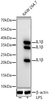
WB
Western blot analysis of lysates from RAW 264.7 cells using IL1β Rabbit mAb at 1:1000 dilution incubated overnight at 4℃. Raw264.7 cells were treated by LPS (1μg/ml) at 37℃ for 8 hours. Secondary antibody: HRP-conjugated Goat anti-Rabbit IgG (H+L) at 1:10000 dilution. Lysates/proteins: 30 μg per lane. Blocking buffer: 3 % nonfat dry milk in TBST. Detection: ECL Basic Kit. Exposure time: 90s.ICC/IF
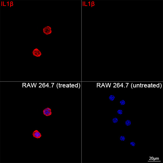
Confocal imaging of RAW 264.7 cells (treated with BFA and LPS) and RAW 264.7 cells (untreated) using IL1β Rabbit mAb (dilution 1:200) followed by a further incubation with Cy3 Goat Anti-Rabbit IgG (H+L) (dilution 1:500) (Red). DAPI was used for nuclear staining (Blue). Objective: 100x.| Product Name | IL1β Rabbit mAb |
|---|---|
| Antibody Type | Primary Antibodies |
| Immunogen | Recombinant fusion protein containing a sequence corresponding to amino acids 118-269 of mouse IL1β (NP_032387.1). |
| Clonality | monoclonal |
|---|---|
| Isotype | IgG |
| Host Species | Rabbit |
| Tested Applications | ICC/IFWB |
| WB:1:1000-1:5000 ICC/IF:1:100-1:400 |
|
| Species Reactivity | Mouse |
| Concentration | 1mg/ml |
| Purification | Affinity purified |
| Gene Symbol | Il1b |
|---|---|
| Gene Synonyms | Il-1b IL-1beta |
| Gene Full Name | interleukin 1 beta |
| Gene Summary | The protein encoded by this gene is a member of the interleukin 1 cytokine family. This cytokine is produced by activated macrophages as a proprotein, which is proteolytically processed to its active form by caspase 1. The encoded protein plays a role in thymocyte proliferation and is involved in the inflammatory response. [provided by RefSeq, Aug 2015] |
| Molecular Weight(MW) | 19kDa/17kDa/31kDa |
| Cellular Localization | cytosol, extracellular region, extracellular space, lysosome. |
WB
Western blot analysis of lysates from RAW 264.7 cells using IL1β Rabbit mAb at 1:1000 dilution incubated overnight at 4℃. Raw264.7 cells were treated by LPS (1μg/ml) at 37℃ for 8 hours. Secondary antibody: HRP-conjugated Goat anti-Rabbit IgG (H+L) at 1:10000 dilution. Lysates/proteins: 30 μg per lane. Blocking buffer: 3 % nonfat dry milk in TBST. Detection: ECL Basic Kit. Exposure time: 90s.
ICC/IF
Confocal imaging of RAW 264.7 cells (treated with BFA and LPS) and RAW 264.7 cells (untreated) using IL1β Rabbit mAb (dilution 1:200) followed by a further incubation with Cy3 Goat Anti-Rabbit IgG (H+L) (dilution 1:500) (Red). DAPI was used for nuclear staining (Blue). Objective: 100x.| Application Notes | WB:1:1000-1:5000 ICC/IF:1:100-1:400 |
|---|
| Form | Liquid |
|---|---|
| Storage Instructions | Store at -20℃. Avoid freeze / thaw cycles. |
| Storage Buffer | Buffer: PBS with 0.05% proclin300, 0.05% BSA, 50% glycerol, pH7.3. |


 Data sheet for OM644085