Application
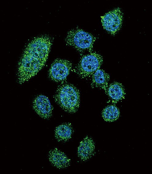
Confocal immunofluorescent analysis of IRF9 Antibody with Hela cell followed by Alexa Fluor 488-conjugated goat anti-rabbit lgG (green).DAPI was used to stain the cell nuclear (blue).Application
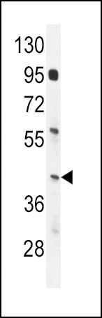
Western blot analysis in mouse spleen tissue lysates (35ug/lane).Application
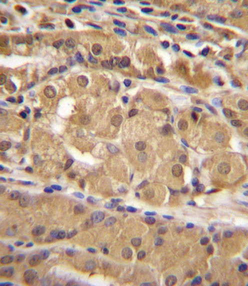
IRF9 Antibody immunohistochemistry analysis in formalin fixed and paraffin embedded human stomach tissue followed by peroxidase conjugation of the secondary antibody and DAB staining.Application
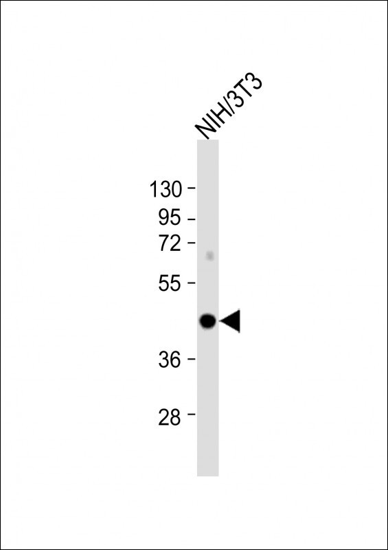
Western Blot at 1:2000 dilution + NIH/3T3 whole cell lysate Lysates/proteins at 20 ug per lane.| Product Name | IRF9 Antibody |
|---|---|
| Antibody Type | Primary Antibodies |
| Product description | IRF9 is a transcription regulatory factor that mediates signaling by type I IFNs (IFN-alpha and IFN-beta). Following type I IFN binding to cell surface receptors, Jak kinases (TYK2 and JAK1) are activated, leading to tyrosine phosphorylation of STAT1 and STAT2. The phosphorylated STATs dimerize, associate with IRF9/ISGF3G to form a complex termed ISGF3 transcription factor, that enters the nucleus. ISGF3 binds to the IFN stimulated response element (ISRE) to activate the transcription of interferon stimulated genes, which drive the cell in an antiviral state.1) Maiwald, T., et al. FEBS J. 277(22):4741-4754(2010) |
| Immunogen | This IRF9 antibody is generated from rabbits immunized with a KLH conjugated synthetic peptide between 75-104 amino acids from the N-terminal region of human IRF9. |
| Clonality | Polyclonal |
|---|---|
| Isotype | Ig |
| Host Species | Rabbit |
| Tested Applications | IFIHC-PWB |
| For WB starting dilution is: 1:1000 For IHC-P starting dilution is: 1:10~50 For IF starting dilution is: 1:10~50 | |
| Species Reactivity | Mouse |
| Concentration | 1mg/ml |
| Purification | Affinity purified |
| Gene Symbol | IRF9 |
|---|---|
| Alternative Names | Interferon regulatory factor 9 IRF-9 IFN-alpha-responsive transcription factor subunit ISGF3 p48 subunit Interferon-stimulated gene factor 3 gamma ISGF-3 gamma Transcriptional regulator ISGF3 subunit gamma IRF9 ISGF3G |
| Molecular Weight(MW) | 44 kDa |
Application
Confocal immunofluorescent analysis of IRF9 Antibody with Hela cell followed by Alexa Fluor 488-conjugated goat anti-rabbit lgG (green).DAPI was used to stain the cell nuclear (blue).
Application
Western blot analysis in mouse spleen tissue lysates (35ug/lane).
Application
IRF9 Antibody immunohistochemistry analysis in formalin fixed and paraffin embedded human stomach tissue followed by peroxidase conjugation of the secondary antibody and DAB staining.
Application
Western Blot at 1:2000 dilution + NIH/3T3 whole cell lysate Lysates/proteins at 20 ug per lane.| Application Notes | For WB starting dilution is: 1:1000 For IHC-P starting dilution is: 1:10~50 For IF starting dilution is: 1:10~50 |
|---|
| Form | Liquid |
|---|---|
| Storage Instructions | Store at 4˚C for three months and -20˚C, stable for up to one year. As with all antibodies care should be taken to avoid repeated freeze thaw cycles. Antibodies should not be exposed to prolonged high temperatures. |
| Storage Buffer | Supplied in PBS with 0.09% (W/V) sodium azide. |


