Application
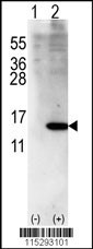
Western blot analysis of ISG15 using rabbit polyclonal ISG15 Antibody (Center R87) using 293 cell lysates (2 ug/lane) either nontransfected (Lane 1) or transiently transfected with ISG15 gene (Lane 2).| Product Name | ISG15 Antibody |
|---|---|
| Antibody Type | Primary Antibodies |
| Antigen Alias | Ubiquitin-like protein ISG15, Interferon-induced 15 kDa protein, Interferon-induced 17 kDa protein, IP17, Ubiquitin cross-reactive protein, hUCRP, ISG15, G1P2, UCRP |
| Product description | ISG15 is secreted from monocytes in response to type I interferons and causes natural killer (NK)-cell proliferation and an augmentation of non-MCH (major histocompatibility complex)-restricted cytotoxicity. Synthesis is stimulated by IFN-alpha or IFN-beta or IFN-omega , but not IFN-gamma . ISG15 expression is also induced by overexpression of interferon regulatory factors that participate in transcriptional regulation of IFN genes, and by influenza B virus. ISG15 is secreted also by cell lines of monocyte, T-lymphocyte, B-lymphocyte, human fibroblasts, and epithelial origins. The induction of terminal differentiation in human melanoma cells is associated with alterations in ISG15 expression. Enhancement of NK cell proliferation, augmentation of non-major histocompatibility complex-restricted cytotoxicity, and induction of IFN-gamma from T cells identify ISG15 as a member of the cytokine cascade and suggest that it may be responsible for amplifying and directing some of the immunomodulatory effects of IFN-alpha or IFN-beta. ISG15 has has also been shown to function intracellularly as a ubiquitin homolog.1) Padovan, E., et al., Cancer Res. 62(12):3453-3458 (2002). |
| Immunogen | This ISG15 antibody is generated from rabbits immunized with a KLH conjugated synthetic peptide between 72-99 amino acids from the Central region of human ISG15. |
| Clonality | Polyclonal |
|---|---|
| Isotype | Ig |
| Host Species | Rabbit |
| Tested Applications | WB |
| For WB starting dilution is: 1:1000: |
|
| Species Reactivity | Human |
| Concentration | 1mg/ml |
| Gene Symbol | ISG15 |
|---|---|
| Alternative Names | Ubiquitin-like protein ISG15 Interferon-induced 15 kDa protein Interferon-induced 17 kDa protein IP17 Ubiquitin cross-reactive protein hUCRP ISG15 G1P2 UCRP |
| Molecular Weight(MW) | 18 kDa |
Application
Western blot analysis of ISG15 using rabbit polyclonal ISG15 Antibody (Center R87) using 293 cell lysates (2 ug/lane) either nontransfected (Lane 1) or transiently transfected with ISG15 gene (Lane 2).| Application Notes | For WB starting dilution is: 1:1000: |
|---|
| Form | Liquid |
|---|---|
| Storage Instructions | Store at 4˚C for three months and -20˚C, stable for up to one year. As with all antibodies care should be taken to avoid repeated freeze thaw cycles. Antibodies should not be exposed to prolonged high temperatures. |
| Storage Buffer | Supplied in PBS with 0.09% (W/V) sodium azide. |


