Application
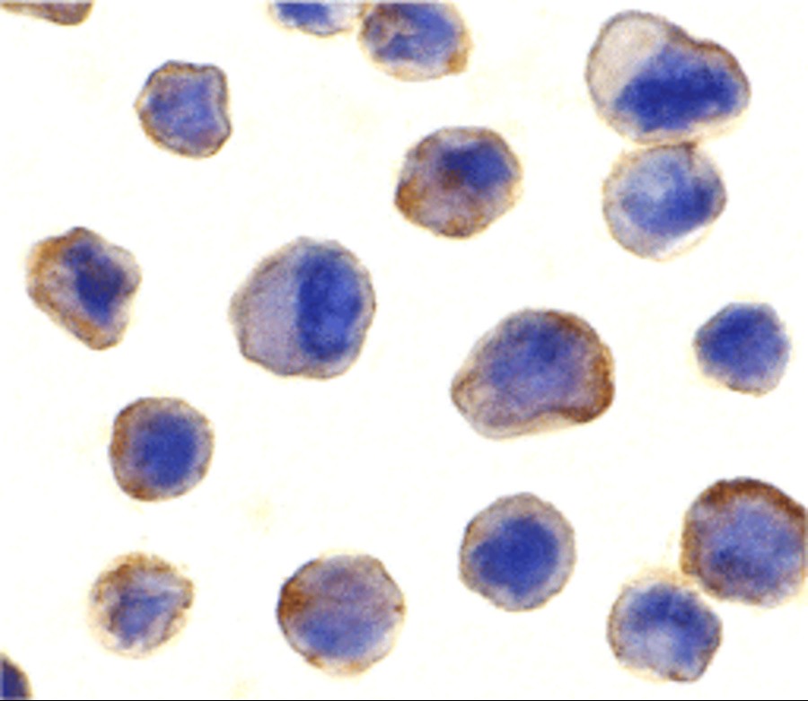
Immunocytochemistry of MADD in human spleen tissue with MADD antibody at 10 μg/mL.Application
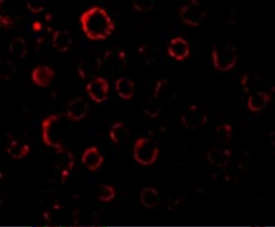
Immunofluorescence of MADD in Hela cells with MADD antibody at 20 μg/mL.| Product Name | MADD Antibody |
|---|---|
| Antibody Type | Primary Antibodies |
| Antigen Alias | MADD Antibody: DENN, IG20, RAB3GEP, DENN, KIAA0358, MAP kinase-activating death domain protein, Differentially expressed in normal and neoplastic cells |
| Product description | MADD Antibody: MAP kinase-activating death domain protein (MADD) was initially identified as the type 1 tumor necrosis factor receptor (TNFR1) associated protein though their death domains. Overexpression of MADD activates MAP kinases ERK and JNK and induces the phosphorylation of cytosolic phospholipase A2. MADD shares 98% identity with DENN (for differentially expressed in neoplastic vs. normal cells), which was recently identified as a substrate for c-jun N-terminal kinase 3 (JNK3). MADD has greater than 94% overall identity to a GDP/GTP exchange protein Rab3-GEP. MADD is 87% identical to KIAA0358, a brain protein of unknown function. Identification of MADD as a component of the TNFR1 signaling complex and the similarity between MADD and Rab3-GEP provides a connection between TNFR1 activation and downstream MAP kinase activity through a guanine-nucleotide exchange protein.1) Schievella AR, Chen JH, Graham JR, Lin LL. MADD, a novel death domain protein that interacts with the type 1 tumor necrosis factor receptor and activates mitogen-activated protein kinase. J Biol Chem 1997;272:12069-12075 |
| Immunogen | MADD antibody was raised against a peptide corresponding to amino acids near the carboxy terminus of human MADD. The immunogen is located within the last 50 amino acids of MADD. |
| Clonality | Polyclonal |
|---|---|
| Isotype | IgG |
| Host Species | Rabbit |
| Tested Applications | ICCIFWB |
| MADD antibody can be used for detection of MADD by Western blot at 1 - 2 mg/mL. 200 to 220 kDa bands should be detected. Antibody can also be used for immunocytochemistry starting at 10 μg/mL. For immunofluorescence start at 20 μg/mL.: |
|
| Species Reactivity | MouseRat |
| Concentration | 1mg/ml |
| Purification | Affinity purified |
| Gene Symbol | MADD |
|---|---|
| Alternative Names | MADD Antibody: DENN IG20 RAB3GEP DENN KIAA0358 MAP kinase-activating death domain protein Differentially expressed in normal and neoplastic cells |
| Molecular Weight(MW) | 200 to 220 kDa |
| Sequence Similarities | 4) Cat. No. 17-001 - HeLa Cell Slide |
Application
Immunocytochemistry of MADD in human spleen tissue with MADD antibody at 10 μg/mL.
Application
Immunofluorescence of MADD in Hela cells with MADD antibody at 20 μg/mL.| Positive Control | 1) Cat. No. 1201 - HeLa Cell Lysate |
|---|---|
| Application Notes | MADD antibody can be used for detection of MADD by Western blot at 1 - 2 mg/mL. 200 to 220 kDa bands should be detected. Antibody can also be used for immunocytochemistry starting at 10 μg/mL. For immunofluorescence start at 20 μg/mL.: |
| Form | Liquid |
|---|---|
| Storage Instructions | MADD antibody can be stored at 4˚C for three months and -20˚C, stable for up to one year. As with all antibodies care should be taken to avoid repeated freeze thaw cycles. Antibodies should not be exposed to prolonged high temperatures. |
| Storage Buffer | MADD Antibody is supplied in PBS containing 0.02% sodium azide. |


